. 2013 Dec 12;39(6):1095-107.
doi: 10.1016/j.immuni.2013.11.003. Epub 2013 Nov 27.

Neonatal Fc receptor expression in dendritic cells mediates protective immunity against colorectal cancer

Affiliations

Neonatal Fc receptor expression in dendritic cells mediates protective immunity against colorectal cancer

Kristi Baker et al. Immunity. .

Abstract

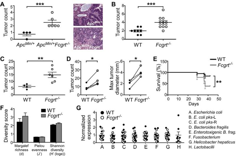
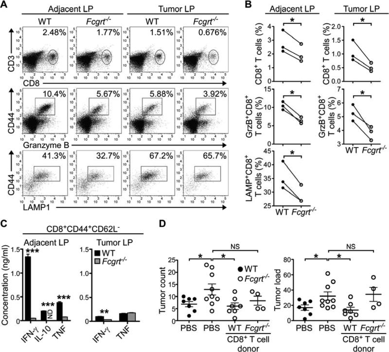
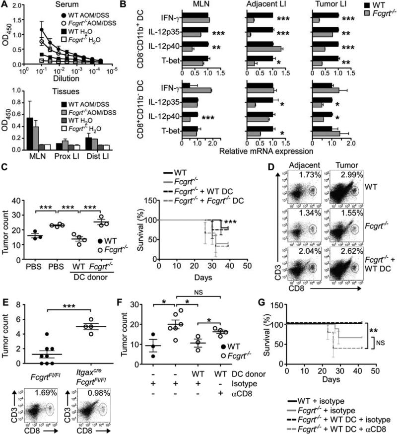
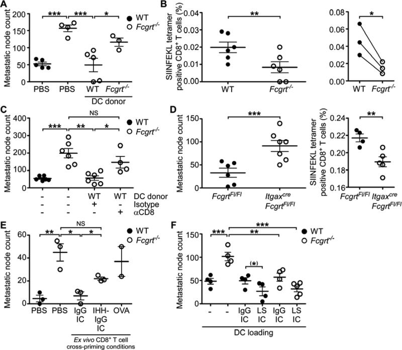
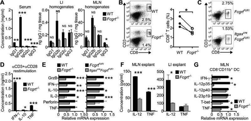
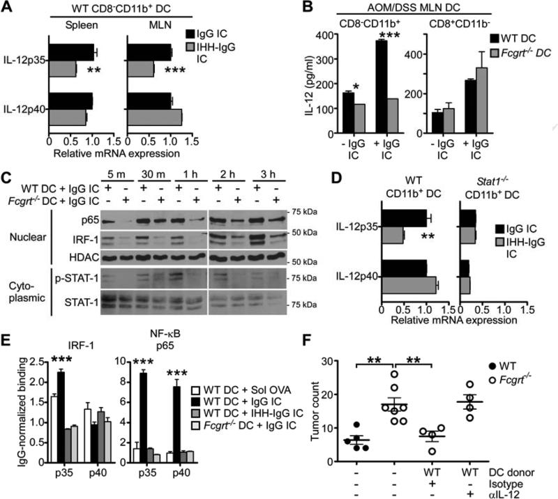
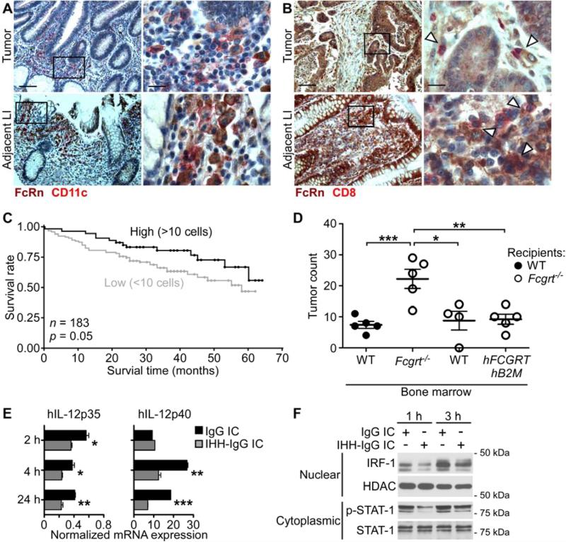
Cancers arising in mucosal tissues account for a disproportionately large fraction of malignancies. Immunoglobulin G (IgG) and the neonatal Fc receptor for IgG (FcRn) have an important function in the mucosal immune system that we have now shown extends to the induction of CD8(+) T cell-mediated antitumor immunity. We demonstrate that FcRn within dendritic cells (DCs) was critical for homeostatic activation of mucosal CD8(+) T cells that drove protection against the development of colorectal cancers and lung metastases. FcRn-mediated tumor protection was driven by DCs activation of endogenous tumor-reactive CD8(+) T cells via the cross-presentation of IgG complexed antigens (IgG IC), as well as the induction of cytotoxicity-promoting cytokine secretion, particularly interleukin-12, both of which were independently triggered by the FcRn-IgG IC interaction in murine and human DCs. FcRn thus has a primary role within mucosal tissues in activating local immune responses that are critical for priming efficient anti-tumor immunosurveillance.


Figures

Figure 1
Figure 1. FcRn protects against the development of colorectal cancer through a mechanism independent of intestinal microbiota
(A) Large intestine (LI) tumor incidence at 5 months of age and representative tumor histology in ApcMin/+ and ApcMin/+Fcgrt−/− mice. Scale bar = 100 μm. (B) Tumor incidence in WT and Fcgrt−/− littermates treated with 8 doses of azoxymethane (AOM). (C) Tumor incidence in AOM/DSS-treated WT and Fcgrt−/− littermates. (D) Tumor incidence and maximum tumor diameter in WT and Fcgrt−/− littermates in each of four independent experiments with n ≥ 3 mice per group per experiment. (E) Percent survival of WT and Fcgrt−/− littermates treated with AOM/DSS. Significance was assessed by Logrank test. (F) Richness indices of microbiota associated with the distal LI of untreated 8-week old WT and Fcgrt−/− littermates, as revealed by T-RFLP analysis. n = 3-5 mice per group. (G) Abundance of specific microbial species in the distal LI of untreated 7-week old WT andFcgrt−/− littermates as assessed by qPCR. n = 9 mice per group. Representative results of two (A,B,E) or four (D) independent experiments each with n = 4-10 mice per group. All data represent mean ± s.e.m. * p ≤ 0.05, ** p ≤ 0.01, *** p ≤ 0.005. See also Figure S1.
Figure 2
Figure 2. FcRn drives the activation and retention of tumor-reactive cytotoxic CD8+ T cells which confer tumor protection
(A) Frequency of CD8+ T cells in the lamina propria lymphocyte (LPL) fraction of tumor and adjacent LI tissue in WT and Fcgrt−/− littermates (upper panels) following AOM/DSS treatment. Cytotoxic potential of cells within the CD3+CD8+ gate was assessed by intracellular staining for granzyme B (middle panels) or surface staining of LAMP1 (lower panels). (B) Mean CD8+ T cell frequency and cytotoxic potential in WT and Fcgrt−/− mice, as assessed by flow cytometry, in each of three independent experiments. (C) Cytokine secretion of sorted effector CD8+ CD44+CD62L cells from the LP of tumor and adjacent tissue of AOM/DSS treated WT and Fcgrt−/− mice following 24 h restimulation with anti-CD3 and anti-CD28. (D) Tumor incidence and tumor load (sum of the diameters of all tumors) in recipient mice adoptively transferred with CD8+ T cells from WT or Fcgrt−/− AOM/DSS-treated donors. Significance was assessed by Mann-Whitney test. Representative results of three independent experiments with n ≥ 4 mice per group per experiment. All data represent mean ± s.e.m. NS = not significant. ND = not detected. * p ≤ 0.05, ** p ≤ 0.01, *** p ≤ 0.005. See also Figure S2.
Figure 3
Figure 3. CD8CD11b+ DC utilize FcRn to efficiently prime protective anti-tumor CD8+ T cell responses
(A) Tumor antigen-specific IgG in the serum or MLN and LI homogenates of AOM/DSS treated WT or Fcgrt−/− mice. ELISA plates coated with lysates from tumor epithelium were probed with dilutions of serum or tissue homogenates from tumor bearing mice. (B) Transcript profiles of sorted CD8CD11b+ and CD8+CD11b DC subsets isolated from the indicated tissue compartment of AOM/DSS-treated WT and Fcgrt−/− littermates. (C) Tumor incidence and survival in Fcgrt−/− recipients adoptively transferred with DC from the MLN and LP of AOM/DSS-treated WT or Fcgrt−/− donors. Endpoint survival was assessed using a Chi-Squared test. (D) CD8+ T cell frequency in the LI LP following transfer of WT DC to AOM/DSS-treated Fcgrt−/− recipients. (E) Tumor incidence and LI LP CD8+ T cell frequency in ItgaxcreFcgrtFl/Fl mice and their littermate FcgrtFl/Fl controls upon treatment with AOM/DSS. (F-G) Tumor incidence (F) and survival (G) of CD8+ T cell-depleted Fcgrt−/− mice adoptively transferred with WT DC. CD8+ T cells were depleted by chronic i.p. administration of anti-CD8 antibody (or isotype control). Representative results of three (B-E) or two (A,F) independent experiments with n = 3-6 mice per group per experiment. All data represent mean ± s.e.m. NS = not significant. * p ≤ 0.05, ** p ≤ 0.01, *** p ≤ 0.005. See also Figure S3.
Figure 4
Figure 4. FcRn drives the induction of endogenous tumor-reactive CD8+ T cells and can be therapeutically targeted
(A) Incidence of pulmonary metastatic nodules formed by i.v. administered OVA-expressing B16 melanoma cells (OVA-B16) in WT or Fcgrt−/− mice or Fcgrt−/− mice pre-immunized with WT or Fcgrt−/− DC. (B) Frequency of endogenously occurring OVA-specific CD8+ T cells in WT and Fcgrt−/− metastasis-bearing mice. Left panel demonstrates results from individual animals in a single experiment. Right panel shows the results of three independent experiments each with n = 3-6 mice per group. (C) Frequency of pulmonary metastases from mice treated as in (A) and given either a CD8+ T cell-depleting antibody or isotype control. (D) Frequency of pulmonary metastatic nodules and OVA-specific CD8+ T cells in the lungs of FcgrtFl/Fl and ItgaxcreFcgrtFl/Fl littermates. (E) Incidence of pulmonary metastatic nodules in WT or Fcgrt−/− mice or Fcgrt−/− mice adoptively transferred with OVA-specific CD8+ T cells primed ex vivo by DC loaded with OVA-containing IgG IC, FcRn non-binding IHH-IgG IC or soluble OVA. (F) Incidence of pulmonary nodules in OVA-B16-treated WT and Fcgrt−/− mice pre-immunized with WT DC loaded ex vivo with OVA-containing IC formed with IgG or enhanced FcRn-binding LS-IgG. Representative results of three (A,B,D) or two (C,E,F) independent experiments with n = 3-6 mice per group per experiment. All data represent mean ± s.e.m. NS = not significant. (*) p = 0.09, * p ≤ 0.05, ** p ≤ 0.01, *** p ≤ 0.005. See also Figure S4.
Figure 5
Figure 5. FcRn within DC enables homeostatic CD8+ T cell activation and IL-12 production in the LI
(A) IgG isotype content of the serum and LI or MLN homogenates in untreated WT and Fcgrt−/− littermates. (B) CD8+ T cell frequency of the LI LPL fraction of untreated WT and Fcgrt−/− littermates in a single experiment (left panels) or across three independent experimental repeats (right panel). (C) Frequency of CD8+ T cells in the LPL fraction of FcgrtFl/Fl and ItgaxcreFcgrtFl/Fl littermates. (D) Cytokine secretion by CD8+ T cells sorted from LI LP of untreated WT and Fcgrt−/− mice following 24 h restimulation with anti-CD3 and anti-CD28. (E) Transcript profiles of CD8+ T cells sorted from LI LP of untreated littermate control mice. (F) Cytokine secretion from 24 h tissue explant cultures of the MLN and LI of untreated WT and Fcgrt−/− mice. (G) Transcript profiles of sorted CD8CD11b+ DC from the MLN of untreated littermates. Representative results of three independent experiments with n = 3-5 mice per group per experiment. All data represent mean ± s.e.m. NS = not significant. * p ≤ 0.05, ** p ≤ 0.01, *** p ≤ 0.005. See also Figure S5.
Figure 6
Figure 6. IgG IC ligation of FcRn in CD8CD11b+ DC induced IL-12 production via activation of a signaling cascade
(A) Induction of IL-12p35 upon ex vivo stimulation of WT CD8CD11b+ DC from the spleen or MLN with IgG IC or FcRn non-binding IHH-IgG IC for 6 h. (B) IL-12 secretion after 24 h IgG IC stimulation of CD8CD11b+ and CD8+CD11b DC sorted from the MLN of AOM/DSS-treated WT and Fcgrt−/− mice. (C) Phosphorylation of STAT-1 and nuclear translocation of IRF-1 and NF-κB p65 upon IgG IC stimulation of DC isolated from WT or Fcgrt−/− mice. (D) IL-12 transcript production by WT or Stat-1−/− CD8CD11b+ DC following stimulation with IgG or IHH-IgG IC for 6 h. (E) Binding of IRF-1 and NF-kB p65 to the promoters of IL-12p35 and IL-12p40 upon stimulation of WT or Fcgrt−/− DC with IgG IC or IHH-IgG IC for 4 h. (F) Tumor incidence in mice adoptively transferred with WT DC and treated with a neutralizing anti-IL-12 antibody or isotype control. Representative results of three (A-E) or one (F) independent experiments with n = 3-7 mice per group per experiment. All data represent mean ± s.e.m. * p ≤ 0.05, ** p ≤ 0.01, *** p ≤ 0.005. See also Figure S6.
Figure 7
Figure 7. FcRn expressing DC predict survival in human CRC and secrete IL-12 upon FcRn stimulation
(A) Double immunohistochemical staining of FcRn+CD11c+ DC in the stroma of CRC (upper panels) and CRC-adjacent normal LI (lower panels). FcRn = brown, CD11c = red. Scale bar left panels = 100 μm. Scale bar right panels = 20 μm. (B) Colocalization of FcRn+ DC (brown) and CD8+ T cells (red) in stroma of CRC (upper panels) and CRC-adjacent normal LI (lower panels). Arrowheads indicate areas of colocalization. (C) Kaplan Meier survival curves of 183 patients with high (≥10 per core) and low (≤ 10 per core) tumor infiltration by CD11c+FcRn+ cells. (D) Incidence of tumors in chimeric mice treated with AOM/DSS. WT recipients were reconstituted with WT bone marrow. Fcgrt−/− recipients were reconstituted with Fcgrt−/−, WT or hFCGRT-hB2M-mFcgrt−/− bone marrow. Representative result of two independent experiments with n = 4-5 mice per group per experiment. (E) hIL-12p35 and hIL-12p40 transcript expression in hMoDC upon stimulation with FcRn-binding (IgG IC) or FcRn non-binding (IHH-IgG IC) immune complexes. (F) Nuclear translocation of IRF-1 and phosphorylation of STAT-1 in hMoDC upon stimulation with IgG IC or IHH-IgG IC. Data in panels A-B are representative of a total of 50 matched CRC and adjacent normal LI pairs. Data in panels E-F are representative of six donors processed in pairs in each of three independent experiments. All data represent mean ± s.e.m. * p ≤ 0.05, ** p ≤ 0.01, *** p ≤ 0.005. See also Figure S7.

References

    1. Antonios D, Rousseau P, Larange A, Kerdine-Romer S, Pallardy M. Mechanisms of IL-12 Synthesis by Human Dendritic Cells Treated with the Chemical Sensitizer NiSO4. J. Immunol. 2010;185:89–98. - PubMed
    1. Aoki K, Tamai Y, Horiike S, Oshima M, Taketo MM. Colonic polyposis caused by mTOR-mediated chromosomal instability in Apc+/delta716 Cdx2+/− compound mutant mice. Nat. Genet. 2003;35:323–330. - PubMed
    1. Arthur JC, Jobin C. The struggle within: Microbial influences on colorectal cancer. Inflamm. Bowel Dis. 2011;17:396–409. - PMC - PubMed
    1. Arthur JC, Perez-Chanona E, Muhlbauer M, Tomkovich S, Uronis JM, Fan TJ, Campbell BJ, Abujamel T, Dogan B, Rogers AB, et al. Intestinal inflammation targets cancer-inducing activity of the microbiota. Science. 2012;338:120–123. - PMC - PubMed
    1. Auer IO, Grosch L, Hardorfer C, Roder A. Ulcerative colitis specific cytotoxic IgG-autoantibodies against colonic epithelial cancer cells. Gut. 1988;29:1639–1647. - PMC - PubMed

Publication types

Substances