Review
. 2014 Sep 15:739:74-82.
doi: 10.1016/j.ejphar.2013.10.070. Epub 2013 Nov 28.

FK506-binding protein 1b/12.6: a key to aging-related hippocampal Ca2+ dysregulation?

Affiliations
Review

FK506-binding protein 1b/12.6: a key to aging-related hippocampal Ca2+ dysregulation?

J C Gant et al. Eur J Pharmacol. .

Abstract

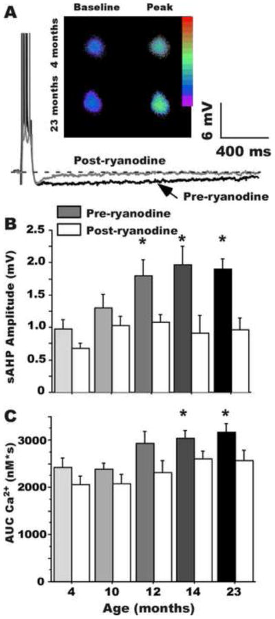
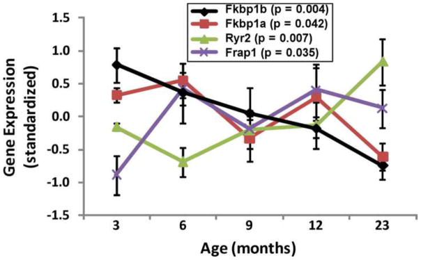
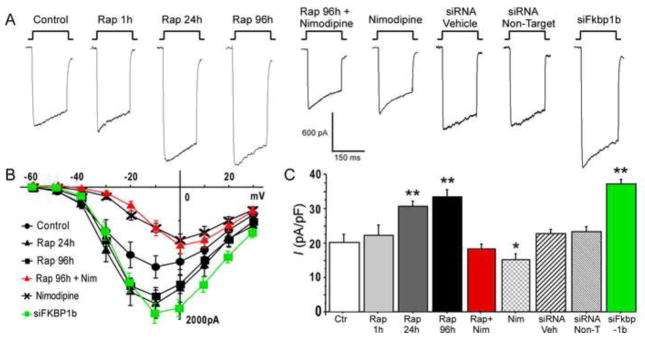
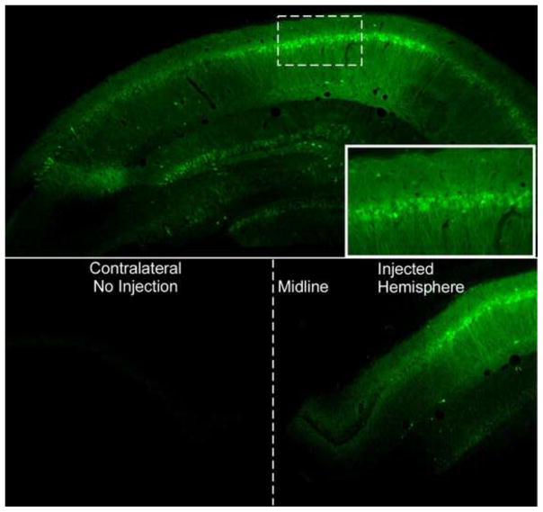
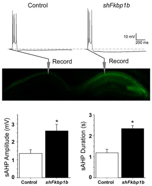
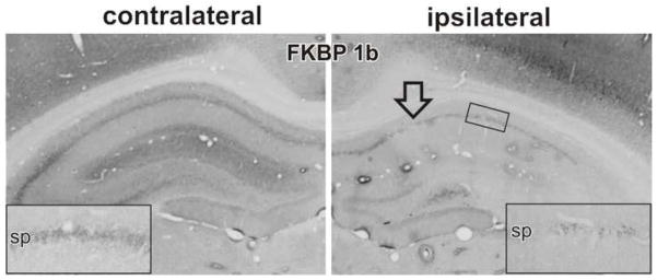
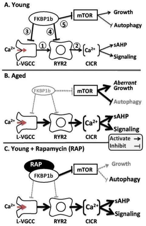
It has been recognized for some time that the Ca(2+)-dependent slow afterhyperpolarization (sAHP) is larger in hippocampal neurons of aged compared with young animals. In addition, extensive studies since have shown that other Ca(2+)-mediated electrophysiological responses are increased in hippocampus with aging, including Ca(2+) transients, L-type voltage-gated Ca(2+) channel activity, Ca(2+) spike duration and action potential accommodation. Elevated Ca(2+)-induced Ca(2+) release from ryanodine receptors (RyRs) appears to drive amplification of the Ca(2+) responses. Components of this Ca(2+) dysregulation phenotype correlate with deficits in cognitive function and plasticity, indicating they may play critical roles in aging-related impairment of brain function. However, the molecular mechanisms underlying aging-related Ca(2+) dysregulation are not well understood. FK506-binding proteins 1a and 1b (FKBP1a/1b, also known as FKBP12/12.6) are immunophilin proteins that bind the immunosuppressant drugs FK506 and rapamycin. In muscle cells, FKBP1a/1b also bind RyRs and inhibits Ca(2+)-induced Ca(2+) release, but it is not clear whether FKBPs act similarly in brain cells. Recently, we found that selectively disrupting hippocampal FKBP1b function in young rats, either by microinjecting adeno-associated viral vectors expressing siRNA, or by treatment with rapamycin, increases the sAHP and recapitulates much of the hippocampal Ca(2+) dysregulation phenotype. Moreover, in microarray studies, we found FKBP1b gene expression was downregulated in hippocampus of aging rats and early-stage Alzheimer's disease subjects. These results suggest the novel hypothesis that declining FKBP function is a key factor in aging-related Ca(2+) dysregulation in the brain and point to potential new therapeutic targets for counteracting unhealthy brain aging.

Keywords: Aging; Calcium; FKBP1b; Ryanodine receptor.


Figures

Figure 1
Figure 1. The AHP and intracellular Ca2+ are altered by aging
A: Example of the sAHP in hippocampal neurons from a young and an aging rat, showing the increased AHP magnitude with aging (darker trace); also shown are examples of the increase in intracellular Ca2+ transients (inset). Age-related increases in sAHP amplitude (B) and Ca2+ transients (C) begin by 12 months-of-age and are blocked by ryanodine, indicating the increases depend on CICR from RyRs (* p < .05). (From Gant et al., 2006)
Figure 2
Figure 2. Age-dependent changes in expression of Fkbp1b, Fkbp1a, Ryr2, and Frap1/mTOR mRNA
(From data in Kadish et al., 2009)
Figure 3
Figure 3. Knockdown of FKBP1b or treatment with rapamycin (Rap) increased Ca2+ channel current in cultured hippocampal neurons
A, Representative whole-cell patch-clamp current traces from each experimental condition; B, Mean current/voltage (I/V) relationships for 6 of the 9 experimental conditions. The I–V curves for Rap 1h, siRNA Veh and siRNA Non-Target were not different from the control condition and are omitted for illustrative clarity. Exposure to siFkbp1b for 96 h or to rapamycin for 24/96 h induced enhancement of VGCC current. Treatment with siFkbp1b or with rapamycin altered the amplitude but not the voltage dependence of Ca2+ current, whereas nimodipine (Nim) shifted peak current to more positive voltage; C, Means +/− S.E.M. of peak Ca2+ current density for the 9 conditions. Asterisks indicate significant differences from the control condition (* p < 0.05 and **p < 0.0001). n = control (18), Rap 1h (4), Rap 24h (20), Rap 96h (20), Rap + Nim (10), Nim (12), siRNA Veh (23), siRNA Non-Target (19), siFkbp1b (23). (From Gant et al., 2011)
Figure 4
Figure 4. Green Flourescent Protein (GFP) expression in the hippocampus
A: GFP expression in the hippocampus following infusion with serotype 2.1 AAV expressing GFP. Note the high level of expression in the pyramidal cells corresponding to the CA regions. Solid line box inset is shows close-up of pyramidal cells expressing GFP from the CA1 region (Dotted box). B: Note that GFP expression is only found in the hippocampus that was injected during a unilateral injection. The contralateral side within the same animal contained no cells expressing GFP. Midline of the hemispheres is denoted with a dotted line.
Figure 5
Figure 5. Knockdown of FKBP1b in vivo enhanced the slow AHP
Top, Representative sharp electrode intracellular recordings showing four triggered action potentials, followed by an AHP (dashed line indicates baseline) in CA1 neurons from slices of a control (left) and an AAV–shFkbp1b-injected (right) hippocampus. Middle, Fluorescent imaging in an animal receiving a similar unilateral AAV injection expressing GFP only, showing strong expression in the injected, but not the contralateral hippocampus, and also illustrating general placement of recording pipettes in the pyramidal neuron somal layer (stratum pyramidale) of field CA1. Bottom, sAHP amplitude (left) and duration (right) were significantly increased by FKBP1b knockdown (shFkbp1b injection) (*p < 0.0025 for either variable, t test; n = 17 neurons per group). (From Gant et al., 2011)
Figure 6
Figure 6. shFkbp1b mediated knockdown of Fkbp1b validated at the protein level
In vivo direct hippocampal shFkbp1b injection into one hemisphere (ipsilateral) is compared to the non-injected contralateral hemisphere. To validate knockdown at the protein level, immunohistochemical staining of FKBP11b/12.6 was performed. Arrow indicates injection site. Inset shows magnified view of CA1 region. (Modified from Gant et al., 2011)
Figure 7
Figure 7. Working model of FKBP1b’s role in aging-related Ca2+ dyshomeostasis
A: In young subjects, neuronal FKBP1b exerts strong tonic inhibitory effects on two pathways, the Ca2+ regulatory pathway and the mTOR signaling pathway. In the Ca2+ regulatory pathway, FKBP1b inhibits cytosolic Ca2+ rises generated by extracellular influx via L-VGCCs (1) and intracellular release from RYRs (2), by inhibiting both channel types directly (3 & 4). In the mTOR pathway, FKBP inhibition of mTOR (5) helps maintain the balance between growth-stimulating and autophagy-suppressing effects of mTOR. B: With aging (mimicked by FKBP1b knockdown), a decline of FKBP1b expression/function leads to weakened inhibition of both pathways, and concomitant increases in both Ca2+ and mTOR signaling. Increased Ca2+ signaling results in dampened neuronal excitability and function, whereas mTOR disinhibition leads to aberrant increases in growth signaling as well as decreased autophagy. C: Rapamycin in young subjects exerts opposite effects on FKBP inhibition in the two pathways, mimicking aging’s effect of weakened FKBP inhibition in the Ca2+ pathway (B), but paradoxically augmenting FKBP inhibition of the mTOR pathway (C). (From Gant et al. 2011)

References

    1. Aitken DH, Meaney MJ. Temporally graded, age-related impairments in spatial memory in the rat. Neurobiol Aging. 1989;10:273–276. - PubMed
    1. Bach ME, Barad M, Son H, Zhuo M, Lu YF, Shih R, Mansuy I, Hawkins RD, Kandel ER. Age-related defects in spatial memory are correlated with defects in the late phase of hippocampal long-term potentiation in vitro and are attenuated by drugs that enhance the cAMP signaling pathway. Proceedings of the National Academy of Sciences of the United States of America. 1999;96:5280–5285. - PMC - PubMed
    1. Berridge MJ. Elementary and global aspects of calcium signalling. J Physiol. 1997;499 (Pt 2):291–306. - PMC - PubMed
    1. Berrout J, Isokawa M. Homeostatic and stimulus-induced coupling of the L-type Ca2+ channel to the ryanodine receptor in the hippocampal neuron in slices. Cell Calcium. 2009;46:30–38. - PMC - PubMed
    1. Blalock EM, Chen KC, Sharrow K, Herman JP, Porter NM, Foster TC, Landfield PW. Gene microarrays in hippocampal aging: statistical profiling identifies novel processes correlated with cognitive impairment. J Neurosci. 2003;23:3807–3819. - PMC - PubMed

Publication types