

Mass spectrometry for the biophysical characterization of therapeutic monoclonal antibodies
- PMID: 24291257
- PMCID: PMC3917544
- DOI: 10.1016/j.febslet.2013.11.027
Mass spectrometry for the biophysical characterization of therapeutic monoclonal antibodies
Abstract
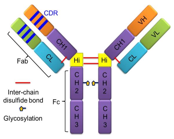
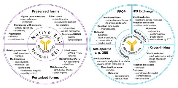
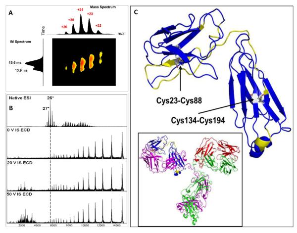
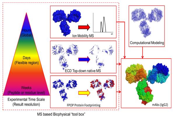
Monoclonal antibodies (mAbs) are powerful therapeutics, and their characterization has drawn considerable attention and urgency. Unlike small-molecule drugs (150-600 Da) that have rigid structures, mAbs (∼150 kDa) are engineered proteins that undergo complicated folding and can exist in a number of low-energy structures, posing a challenge for traditional methods in structural biology. Mass spectrometry (MS)-based biophysical characterization approaches can provide structural information, bringing high sensitivity, fast turnaround, and small sample consumption. This review outlines various MS-based strategies for protein biophysical characterization and then reviews how these strategies provide structural information of mAbs at the protein level (intact or top-down approaches), peptide, and residue level (bottom-up approaches), affording information on higher order structure, aggregation, and the nature of antibody complexes.
Keywords: FPOP; Hydrogen/deuterium exchange; Ion mobility; Mass spectrometry; Monoclonal antibody; Native ESI; Protein footprinting; Top-down and bottom-up.
Copyright © 2013 Federation of European Biochemical Societies. Published by Elsevier B.V. All rights reserved.
Figures
References
-
- Buss NA, Henderson SJ, McFarlane M, Shenton JM, de Haan L. Monoclonal antibody therapeutics: history and future. Curr Opin Pharmacol. 2012;12:615–22. - PubMed
-
- Larrick JW. Conference scene: progress with promising human antibodies. Immunotherapy. 2012;4:257–61. - PubMed
-
- Nelson AL, Dhimolea E, Reichert JM. Development trends for human monoclonal antibody therapeutics. Nat Rev Drug Discov. 2010;9:767–74. - PubMed
-
- Metzger H, Kinet JP. How antibodies work: focus on Fc receptors. FASEB J. 1988;2:3–11. - PubMed
-
- Raghavan M, Bjorkman PJ. Fc receptors and their interactions with immunoglobulins. Annu Rev Cell Dev Biol. 1996;12:181–220. - PubMed
Publication types
MeSH terms
Substances
Grants and funding
LinkOut - more resources
Full Text Sources
Other Literature Sources
