. 2014 Sep 12:276:135-47.
doi: 10.1016/j.neuroscience.2013.11.007. Epub 2013 Nov 28.

Role of myelin plasticity in oscillations and synchrony of neuronal activity

Affiliations

Role of myelin plasticity in oscillations and synchrony of neuronal activity

S Pajevic et al. Neuroscience. .

Abstract

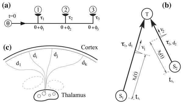
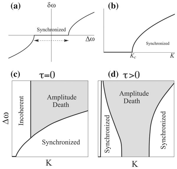
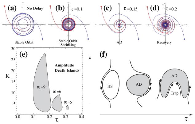
Conduction time is typically ignored in computational models of neural network function. Here we consider the effects of conduction delays on the synchrony of neuronal activity and neural oscillators, and evaluate the consequences of allowing conduction velocity (CV) to be regulated adaptively. We propose that CV variation, mediated by myelin, could provide an important mechanism of activity-dependent nervous system plasticity. Even small changes in CV, resulting from small changes in myelin thickness or nodal structure, could have profound effects on neuronal network function in terms of spike-time arrival, oscillation frequency, oscillator coupling, and propagation of brain waves. For example, a conduction delay of 5ms could change interactions of two coupled oscillators at the upper end of the gamma frequency range (∼100Hz) from constructive to destructive interference; delays smaller than 1ms could change the phase by 30°, significantly affecting signal amplitude. Myelin plasticity, as another form of activity-dependent plasticity, is relevant not only to nervous system development but also to complex information processing tasks that involve coupling and synchrony among different brain rhythms. We use coupled oscillator models with time delays to explore the importance of adaptive time delays and adaptive synaptic strengths. The impairment of activity-dependent myelination and the loss of adaptive time delays may contribute to disorders where hyper- and hypo-synchrony of neuronal firing leads to dysfunction (e.g., dyslexia, schizophrenia, epilepsy).

Keywords: activity-dependent myelination; conduction velocity and delays; coupled oscillators; oscillations; synchronization; white matter plasticity.


Figures

Fig. 1
Fig. 1
(a) Connectivity and synaptic strength cannot fully describe the functioning of a system with time delays and variable propagation speeds. (b) Two neurons, S1 and S2, firing at times t1 and t2, are sending action potentials to a single target neuron along corresponding axons. The spikes propagate at different conduction speeds v1 and v2, according to xi(t) = vi(tti), i = 1, 2, and hence arrive at potentially different times, t1 + d1/v1 versus t2 + d2/v2 unless v1 and v2 are matched to encode a particular time-locked pattern. (c) In recurrent thalamo-cortical resonance large-scale synchronized events between remote brain regions occur. The distances, di, from a given thalamic nuclei to different cortical areas can differ significantly, hence the task of synchronizing distant cortical areas requires the conduction delays to be adjusted.
Fig. 2
Fig. 2
The dynamical state of coupled oscillators as a function of the spread of natural frequencies of these oscillators, Δω, and the coupling strength, K. In the Kuramoto model synchronization occurs for (a) a sufficiently small mismatch Δω, as shown with zero frequency spread, δω, and for (b) a coupling strength that is larger than some critical value (assuming coupling is sufficiently large in (a), and the mismatch is not too large in (b)). A better way to depict dynamic regimes is to use state diagrams, which show more clearly what pair of values (K, Δω) will produce synchronization, or amplitude death for amplitude-coupled models. State diagrams for such a model are shown for (c) no time delays, and (d) when time delay is sufficiently large to affect the dynamics. Note that with the presence of time delay even the oscillators with identical frequencies (Δω = 0) can lead to AD for moderate coupling strength.
Fig. 3
Fig. 3
The dynamical state of coupled oscillators as a function of the coupling strength, K, and the time delay, τ. (a–d) Dynamics of two amplitude-coupled non-linear oscillators, as described by Eq. (6), for K = 5, ω1 = 8, ω2 = 9, and for different values of time lags as indicated. The trajectory of the two oscillators is depicted in a complex plane (containing the information about the amplitude and the phase) with red and blue lines respectively, where the circled dots indicate the starting positions, and plain blue and red dots indicate the positions at time t = 20. These positions end up in a large and stable limit cycle for zero time delay, for virtually any initial conditions as shown in (a). Increasing the time delay in the oscillator interactions leads to a decrease in amplitude (b), and for a range of values of delay it leads to amplitude death, i.e., at the origin of the complex plane, as shown in (c). Further increase of τ recovers the limit cycle, and start of the recovery is shown in (d). This behavior is summarized in (e) using (K,τ) state diagrams, which clearly shows the isolated regions of AD, the death islands, in which activity is quenched after a short period of time. This state diagram is calculated for coupled oscillators with matching frequencies, ω1 = ω2 = ω, and for three different values of ω, as indicated. (f) A sketch of how a dynamical system, in which K and τ can be changed adaptively and are functions of activity, can navigate itself away from the regions with hyper-synchronizability (HS), or amplitude AD regions. Depending on the shape of such regions and the type of dependence of the parameters K and τ on the activity, the system might still end up being trapped and unable to adapt further without entering the “death islands”.

References

    1. Abraham H, Vincze A, Jewgenow I, Beszpremi B, Kravjak A, Gomori E, Seress L. Myelination in the human hippocampal formation form midgestation to adulthood. Int J Dev Neurosci. 2010;28:401–410. - PubMed
    1. Ainsworth M, Lee S, Cunningham MO, Traub RD, Kopell NJ, Whittington MA. Rates and rhythms: a synergistic view of frequency and temporal coding in neuronal networks. Neuron. 2012;75:572–583. - PubMed
    1. Aronson DG, Ermentrout GB, Kopell N. Amplitude response of coupled oscillators. Phys D Nonlinear Phenom. 1990;41:403–449.
    1. Bakiri Y, Burzomato V, Frugier G, Hamilton NB, Karadottir R, Attwell D. Glutamatergic signaling in the brain's white matter. Neuroscience. 2009;158:266–274. - PubMed
    1. Bar-Eli K. On the stability of coupled chemical oscillators. Phys D Nonlinear Phenom. 1985;14:242–252.