. 2014 Feb;7(2):225-32.
doi: 10.1242/dmm.013664. Epub 2013 Nov 28.

Luminal epithelium in endometrial fragments affects their vascularization, growth and morphological development into endometriosis-like lesions in mice

Affiliations

Luminal epithelium in endometrial fragments affects their vascularization, growth and morphological development into endometriosis-like lesions in mice

Dilu Feng et al. Dis Model Mech. 2014 Feb.

Abstract

In endometriosis research, endometriosis-like lesions are usually induced in rodents by transplantation of isolated endometrial tissue fragments to ectopic sites. In the present study, we investigated whether this approach is affected by the cellular composition of the grafts. For this purpose, endometrial tissue fragments covered with luminal epithelium (LE(+)) and without luminal epithelium (LE(-)) were transplanted from transgenic green-fluorescent-protein-positive (GFP(+)) donor mice into the dorsal skinfold chamber of GFP(-) wild-type recipient animals to analyze their vascularization, growth and morphology by means of repetitive intravital fluorescence microscopy, histology and immunohistochemistry during a 14-day observation period. LE(-) fragments developed into typical endometriosis-like lesions with cyst-like dilated endometrial glands and a well-vascularized endometrial stroma. In contrast, LE(+) fragments exhibited a polypoid morphology and a significantly reduced blood perfusion after engraftment, because the luminal epithelium prevented the vascular interconnection with the microvasculature of the surrounding host tissue. This was associated with a markedly decreased growth rate of LE(+) lesions compared with LE(-) lesions. In addition, we found that many GFP(+) microvessels grew outside the LE(-) lesions and developed interconnections to the host microvasculature, indicating that inosculation is an important mechanism in the vascularization process of endometriosis-like lesions. Our findings demonstrate that the luminal epithelium crucially affects the vascularization, growth and morphology of endometriosis-like lesions. Therefore, it is of major importance to standardize the cellular composition of endometrial grafts in order to increase the validity and reliability of pre-clinical rodent studies in endometriosis research.

Keywords: Angiogenesis; Dorsal skinfold chamber; Endometriosis; Endometriotic lesion; Intravital fluorescence microscopy; Luminal epithelium; Morphology; Vascularization.


Figures
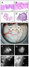
Fig. 1.
Fig. 1.
Dorsal skinfold chamber model of endometriosis. (A,B) H&E-stained sections of the longitudinally opened uterine horns from a C57BL/6-TgN(ACTB-EGFP)1Osb/J donor mouse for the isolation of a LE fragment (A, dotted line) and a LE+ fragment (B, dotted line). The LE+ fragment was excised from the luminal side of the endometrium of an intact uterine horn, which exhibits a layer of myometrium with the underlying perimetrium (B, asterisk). For the isolation of the LE fragment, this layer is first removed (compare A with B) and the fragment is then excised from the exposed basal endometrium (A). (C,D) H&E-stained sections of a LE fragment (C) and a LE+ fragment (D) directly after the isolation procedure. The luminal epithelium covering the LE+ fragment is clearly visible (D, arrows). (E) Observation window of the dorsal skinfold chamber of a C57BL/6 wild-type mouse directly after transplantation of a LE fragment and a LE+ fragment onto the host striated muscle tissue (transplants are indicated by arrows). (F–I) Intravital fluorescent microscopic images of a LE fragment (F,H) and a LE+ fragment (G,I) directly after transplantation into the dorsal skinfold chamber. The fragments can be easily detected in blue light epi-illumination due to their GFP signal. The luminal epithelium of the LE+ fragment is clearly visible at higher magnification (I, arrows). Scale bars: 170 μm (A,B), 90 μm (C,D), 1.3 cm (E), 220 μm (F,G), 20 μm (H,I).
Fig. 2.
Fig. 2.
Vascularization of transplanted endometrial tissue fragments. (A,B) Intravital fluorescent microscopic images of a vascularized LE fragment (A, borders marked by dotted line) and a LE+ fragment (B, borders marked by dotted line) at day 14 after transplantation into the dorsal skinfold chamber of a C57BL/6 wild-type mouse. The microvascular network of the LE fragment exhibits many interconnections to the host microvasculature of the chamber tissue (A), whereas only a few microvessels pierce into the LE+ fragment (B, arrows). Blue light epi-illumination with contrast enhancement by intravascular staining of plasma with 5% FITC-labeled dextran 150,000 (i.v.). Scale bars: 290 μm. (C,D) Functional capillary density (cm/cm2) and number of border crossing vessels (at day 14) of LE fragments (white circles and bar graph, n=8) and LE+ fragments (black circles and bar graph, n=8) after transplantation into dorsal skinfold chambers of C57BL/6 wild-type mice, as assessed by intravital fluorescence microscopy and computer-assisted image analysis. Values are means ± s.e.m. aP<0.05 versus day 0 within each individual group; bP<0.05 versus days 0 and 3 within each individual group; cP<0.05 versus days 0, 3 and 6 within each individual group; *P<0.05 versus LE+ fragments.
Fig. 3.
Fig. 3.
Barrier function of the luminal epithelium during fragment vascularization. (A,B) Intravital fluorescence microscopic images of a LE fragment (A) and a LE+ fragment (B) at day 6 after transplantation into the dorsal skinfold chamber of a C57BL/6 wild-type mouse. (C,D) Higher magnification images of windows in A and B, respectively. The detection of GFP in blue light epi-illumination reveals that many GFP+ microvessels (C, arrows) grow out of the LE fragment. By contrast, in the LE+ fragment microvessels do not pass the luminal epithelium and, thus, do not grow out of the graft (D). Scale bars: 310 μm (A,B), 90 μm (C,D).
Fig. 4.
Fig. 4.
Microhemodynamics of transplanted endometrial tissue fragments. (A–C) Diameter (A), centerline RBC velocity (B) and volumetric blood flow (C) of microvessels in LE fragments (white circles, n=8) and LE+ fragments (black circles, n=8) after transplantation into dorsal skinfold chambers of C57BL/6 wild-type mice, as assessed by intravital fluorescence microscopy and computer-assisted image analysis. Values are means ± s.e.m. aP<0.05 versus day 3 within each individual group; *P<0.05 versus LE+ fragments.
Fig. 5.
Fig. 5.
Size of transplanted endometrial tissue fragments. Size (as a percentage of initial size) of LE fragments (white bar, n=8) and LE+ fragments (black bar, n=8) at day 14 after transplantation into dorsal skinfold chambers of C57BL/6 wild-type mice, as assessed by intravital fluorescence microscopy and computer-assisted image analysis. Values are means ± s.e.m. *P<0.05 versus LE+ fragments.
Fig. 6.
Fig. 6.
Histomorphology and blood vessel origin of transplanted endometrial tissue fragments. (A,B) H&E-stained sections of a LE fragment (A, borders marked by dashed line) and a LE+ fragment (B, borders marked by dashed line) at day 14 after transplantation onto the host striated muscle tissue (arrows) of the dorsal skinfold chamber. The LE fragment has developed into a typical endometriosis-like lesion with cyst-like dilated endometrial glands, which are surrounded by a well-vascularized stroma. In contrast, the lesion originating from the LE+ fragment exhibits a polypoid morphology with less glands and a coverage of luminal epithelium. (C–H) Immunohistochemical analysis of the origin of microvessels within and around a LE fragment (C–E) and a LE+ fragment (F–H) at day 14 after transplantation into the dorsal skinfold chamber of a C57BL/6 wild-type mouse. Histological sections were stained with Hoechst 33342 to identify cell nuclei (C–H, blue), an antibody against CD31 for the detection of endothelial cells (C,F, red) and an antibody against GFP (D,G, green). E displays a merge of C and D, and H a merge of F and G. In contrast to the polypoid lesion originating from the LE+ fragment, the endometriosis-like lesion originating from the LE fragment is surrounded by many GFP+ microvessels (arrows). Scale bars: 75 μm (A,B), 70 μm (C–H).
Fig. 7.
Fig. 7.
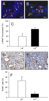
Vessel maturation and proliferating activity of transplanted endometrial tissue fragments. (A,B) Immunohistochemical analysis of vessel maturation within a LE fragment (A) and a LE+ fragment (B) at day 14 after transplantation into the dorsal skinfold chamber of a C57BL/6 wild-type mouse. A and B display merges of histological sections stained with Hoechst 33342 to identify cell nuclei (blue), an antibody against CD31 for the detection of microvessels (red) and an antibody against α-SMA (green). Both fragment types contain a mixture of α-SMA+ mature microvessels (arrows) and α-SMA immature microvessels (arrowheads). (C) α-SMA+ microvessels (as a percentage of the total number of microvessels) within LE fragments (white bar, n=6) and LE+ fragments (black bar, n=4), as assessed by immunohistochemical analysis. (D,E) Immunohistochemical detection of PCNA+ cells (brown) within a LE fragment (D) and a LE+ fragment (E) at day 14 after transplantation into the dorsal skinfold chamber of a C57BL/6 wild-type mouse. (F) PCNA+ cells (as a percentage of the total number of cells) within LE fragments (white bar, n=7) and LE+ fragments (black bar, n=6), as assessed by immunohistochemical analysis. Values are means ± s.e.m. *P<0.05 versus LE+ fragments. Scale bars: 30 μm (A,B), 20 μm (D,E).

References

    1. Baker M., Wayland H. (1974). On-line volume flow rate and velocity profile measurement for blood in microvessels. Microvasc. Res. 7, 131–143 - PubMed
    1. Feng D., Welker S., Körbel C., Rudzitis-Auth J., Menger M. D., Montenarh M., Laschke M. W. (2012). Protein kinase CK2 is a regulator of angiogenesis in endometriotic lesions. Angiogenesis 15, 243–252 - PubMed
    1. Gao X., Yeh Y. C., Outley J., Simon J., Botteman M., Spalding J. (2006). Health-related quality of life burden of women with endometriosis: a literature review. Curr. Med. Res. Opin. 22, 1787–1797 - PubMed
    1. Gipson I. K., Blalock T., Tisdale A., Spurr-Michaud S., Allcorn S., Stavreus-Evers A., Gemzell K. (2008). MUC16 is lost from the uterodome (pinopode) surface of the receptive human endometrium: in vitro evidence that MUC16 is a barrier to trophoblast adherence. Biol. Reprod. 78, 134–142 - PubMed
    1. Groothuis P. G., Nap A. W., Winterhager E., Grümmer R. (2005). Vascular development in endometriosis. Angiogenesis 8, 147–156 - PubMed

Publication types