. 2014 Feb;7(2):215-24.
doi: 10.1242/dmm.013490. Epub 2013 Nov 28.

Hypoxia promotes liver-stage malaria infection in primary human hepatocytes in vitro

Affiliations

Hypoxia promotes liver-stage malaria infection in primary human hepatocytes in vitro

Shengyong Ng et al. Dis Model Mech. 2014 Feb.

Abstract

Homeostasis of mammalian cell function strictly depends on balancing oxygen exposure to maintain energy metabolism without producing excessive reactive oxygen species. In vivo, cells in different tissues are exposed to a wide range of oxygen concentrations, and yet in vitro models almost exclusively expose cultured cells to higher, atmospheric oxygen levels. Existing models of liver-stage malaria that utilize primary human hepatocytes typically exhibit low in vitro infection efficiencies, possibly due to missing microenvironmental support signals. One cue that could influence the infection capacity of cultured human hepatocytes is the dissolved oxygen concentration. We developed a microscale human liver platform comprised of precisely patterned primary human hepatocytes and nonparenchymal cells to model liver-stage malaria, but the oxygen concentrations are typically higher in the in vitro liver platform than anywhere along the hepatic sinusoid. Indeed, we observed that liver-stage Plasmodium parasite development in vivo correlates with hepatic sinusoidal oxygen gradients. Therefore, we hypothesized that in vitro liver-stage malaria infection efficiencies might improve under hypoxia. Using the infection of micropatterned co-cultures with Plasmodium berghei, Plasmodium yoelii or Plasmodium falciparum as a model, we observed that ambient hypoxia resulted in increased survival of exo-erythrocytic forms (EEFs) in hepatocytes and improved parasite development in a subset of surviving EEFs, based on EEF size. Further, the effective cell surface oxygen tensions (pO2) experienced by the hepatocytes, as predicted by a mathematical model, were systematically perturbed by varying culture parameters such as hepatocyte density and height of the medium, uncovering an optimal cell surface pO2 to maximize the number of mature EEFs. Initial mechanistic experiments revealed that treatment of primary human hepatocytes with the hypoxia mimetic, cobalt(II) chloride, as well as a HIF-1α activator, dimethyloxalylglycine, also enhance P. berghei infection, suggesting that the effect of hypoxia on infection is mediated in part by host-dependent HIF-1α mechanisms.

Keywords: Hypoxia; Liver-stage malaria; Primary hepatocytes.


Figures

Fig. 1.
Fig. 1.
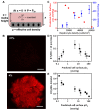
Plasmodium EEF development correlates with hepatic oxygen gradients in vivo. (A) Schematic of liver sinusoid denoting the definition of periportal (PP) EEFs and perivenous (PV) EEFs used for EEF size quantification. (B) 50-μm liver slices were stained with DAPI, and confocal z-stacks were made of GFP-expressing P. yoelii EEFs within 8 hepatocyte lengths of either the portal triad (periportal) or the central vein (perivenous) for which the maximal XY area could be determined within the slice. (C) Maximal XY areas of P. yoelii perivenous or periportal EEFs (as defined for A) at 46 hours post-infection in murine liver; **P<0.01, two-tailed t-test. Scale bar: 50 μm. PV, portal vein; BD, bile duct; HA, hepatic artery; CV, central vein.
Fig. 2.
Fig. 2.
Ambient hypoxia increases liver-stage malaria infection in vitro. (A,B) Ambient hypoxia (4% O2) increases the number of P. berghei and P. yoelii EEFs in PHH MPCCs at 48 hours post-infection. (C,D,G) Ambient hypoxia (black symbols or bars, 4% O2) increases the EEF size distribution of P. berghei and P. yoelii at 48 hours post-infection and P. falciparum at 4 and 6 days post-infection in PHH MPCCs compared with normoxia (white symbols or bars, 21% O2). (E,F,H) Representative immunofluorescence images of P. berghei, P. yoelii EEFs at 48 hours post-infection, and P. falciparum EEFs at 6 days post-infection at either ambient 21% or 4% O2. EEFs were stained for Plasmodium HSP70 (clone 2E6 for P. berghei and P. yoelii, clone 4C9 for P. falciparum). Scale bars: 5 μm. *P<0.05, **P<0.01, ****P<0.0001; two-tailed t-test.
Fig. 3.
Fig. 3.
Optimal pO2 exists for development of mature Plasmodium EEFs. (A) Schematic of steady-state diffusion-reaction model with three parameters that determine cell surface oxygen concentration: atmospheric pO2 (Pair), height of medium and cell density. (B) Validation of effect of atmospheric pO2 on cell surface pO2 by Hypoxyprobe™ staining. Hypoxyprobe™ forms covalent adducts with thiol groups at pO2<10 mmHg. (C) Modulation of cell surface pO2 by varying effective cell density as predicted by the model (red), and Hypoxyprobe™ fluorescence intensity (blue). (D,E) Modulation of cell surface pO2 by simultaneously varying both atmospheric pO2 and effective cell density results (D) in a biphasic relationship between the number of well-developed P. yoelii EEFs and the predicted cell surface pO2 and (E) in a monotonic relationship between the total number of P. yoelii EEFs versus predicted cell surface pO2 in PHH MPCCs at 48 hours post-infection. Scale bars: 100 μm.
Fig. 4.
Fig. 4.
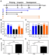
Kinetics of hypoxic treatment alters liver-stage malaria infection in vitro. (A) Schematic of differential hypoxia treatment regimes. (B) Effect of differential hypoxia kinetic regimes on the number of P. berghei EEFs at 48 hours post-infection. (C) Effect of differential hypoxia kinetic regimes on P. berghei EEF sizes at 48 hours post-infection. (D) Effect of ambient hypoxia on P. berghei sporozoite gliding. (E) Effect of ambient hypoxia on P. berghei sporozoite entry into hepatocytes at 3 hours post-infection. **P<0.01, ***P<0.001; one way ANOVA with Tukey’s multiple comparison test.
Fig. 5.
Fig. 5.
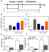
Host HIF-1α induction increases EEF numbers in infected hepatocytes. (A) Schematic of cobalt(II) chloride treatment of PHH MPCCs during infection with P. berghei. (B,C) Effect of cobalt(II) treatment of PHH MPCCs at 21% O2 on (B) the number of P. berghei EEFs at 48 hours post-infection and (C) on the percentage of P. berghei EEFs of >10 μm at 48 hours post-infection; **P<0.01, ***P<0.001, one way ANOVA with Tukey’s multiple comparison test. (D,E) Effect of DMOG treatment of PHH MPCCs at 21% O2 on (D) the numbers of P. berghei EEFs and (E) the number of P. yoelii EEFs at 48 hours post-infection; *P<0.05, two-tailed t-test.

References

    1. Allen J. W., Bhatia S. N. (2003). Formation of steady-state oxygen gradients in vitro: application to liver zonation. Biotechnol. Bioeng. 82, 253–262 - PubMed
    1. Allen J. W., Khetani S. R., Bhatia S. N. (2005). In vitro zonation and toxicity in a hepatocyte bioreactor. Toxicol. Sci. 84, 110–119 - PubMed
    1. Arrais-Silva W. W., Pinto E. F., Rossi-Bergmann B., Giorgio S. (2006). Hyperbaric oxygen therapy reduces the size of Leishmania amazonensis-induced soft tissue lesions in mice. Acta Trop. 98, 130–136 - PubMed
    1. Arteel G. E., Thurman R. G., Yates J. M., Raleigh J. A. (1995). Evidence that hypoxia markers detect oxygen gradients in liver: pimonidazole and retrograde perfusion of rat liver. Br. J. Cancer 72, 889–895 - PMC - PubMed
    1. Bhatia S. N., Toner M., Foy B. D., Rotem A., O’Neil K. M., Tompkins R. G., Yarmush M. L. (1996). Zonal liver cell heterogeneity: effects of oxygen on metabolic functions of hepatocytes. J. Cell. Eng. 1, 125–135

Publication types

MeSH terms

Substances