. 2014 Apr;127(4):523-38.
doi: 10.1007/s00401-013-1220-8. Epub 2013 Nov 30.

Early loss of oligodendrocytes in human and experimental neuromyelitis optica lesions

Affiliations

Early loss of oligodendrocytes in human and experimental neuromyelitis optica lesions

Claudia Wrzos et al. Acta Neuropathol. 2014 Apr.

Abstract

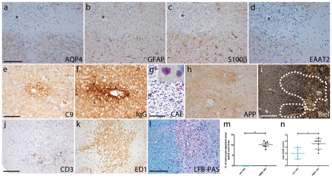
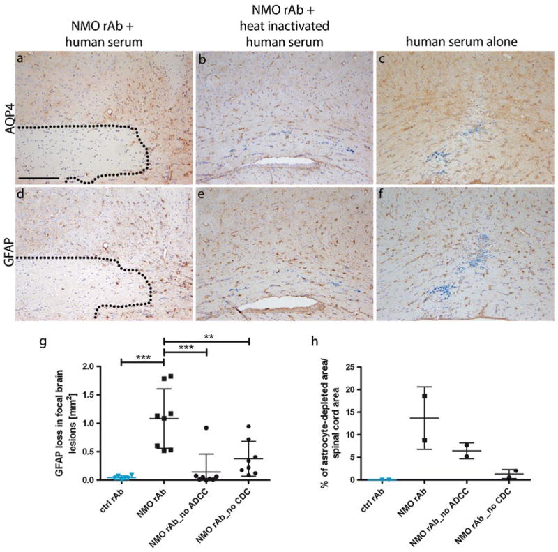
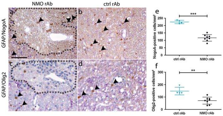
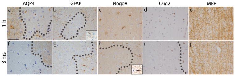
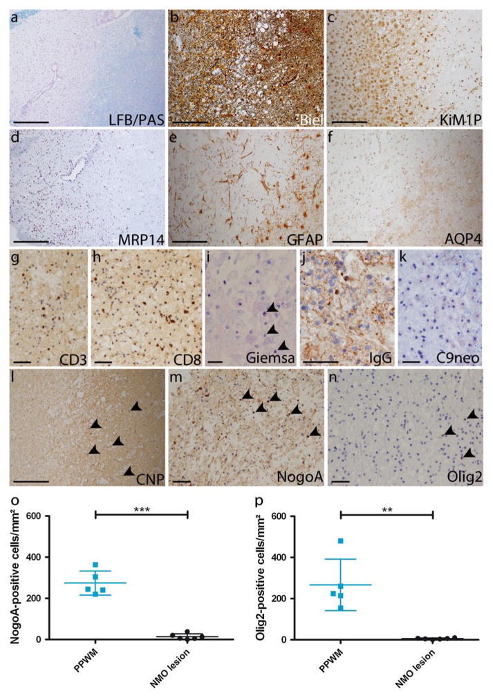
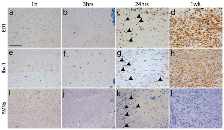
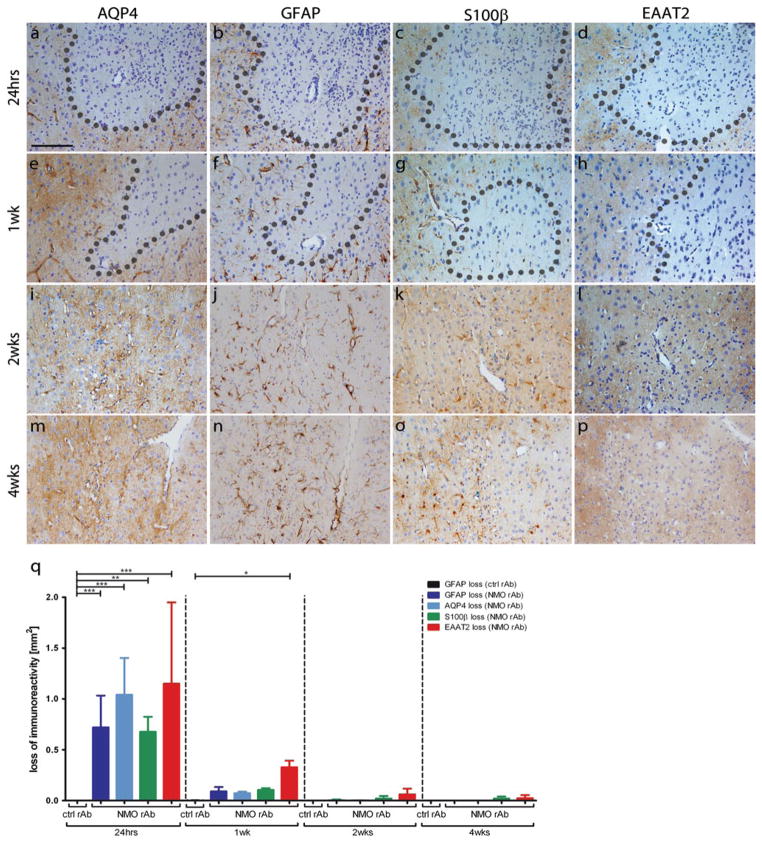
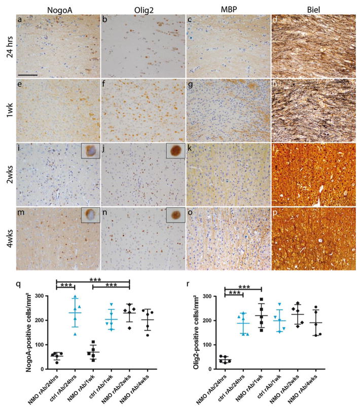
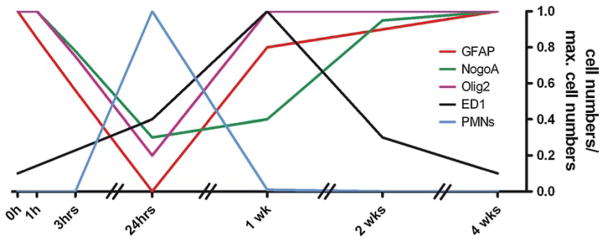
Neuromyelitis optica (NMO) is a chronic, mostly relapsing inflammatory demyelinating disease of the CNS characterized by serum anti-aquaporin 4 (AQP4) antibodies in the majority of patients. Anti-AQP4 antibodies derived from NMO patients target and deplete astrocytes in experimental models when co-injected with complement. However, the time course and mechanisms of oligodendrocyte loss and demyelination and the fate of oligodendrocyte precursor cells (OPC) have not been examined in detail. Also, no studies regarding astrocyte repopulation of experimental NMO lesions have been reported. We utilized two rat models using either systemic transfer or focal intracerebral injection of recombinant human anti-AQP4 antibodies to generate NMO-like lesions. Time-course experiments were performed to examine oligodendroglial and astroglial damage and repair. In addition, oligodendrocyte pathology was studied in early human NMO lesions. Apart from early complement-mediated astrocyte destruction, we observed a prominent, very early loss of oligodendrocytes and oligodendrocyte precursor cells (OPCs) as well as a delayed loss of myelin. Astrocyte repopulation of focal NMO lesions was already substantial after 1 week. Olig2-positive OPCs reappeared before NogoA-positive, mature oligodendrocytes. Thus, using two experimental models that closely mimic the human disease, our study demonstrates that oligodendrocyte and OPC loss is an extremely early feature in the formation of human and experimental NMO lesions and leads to subsequent, delayed demyelination, highlighting an important difference in the pathogenesis of MS and NMO.


Conflict of interest statement

Conflict of interest The authors declare that they have no conflict of interest.

Figures

Fig. 1
Fig. 1
Astrocyte depletion by NMO rAb in spinal lesions in EAE rats. Loss of astrocytic proteins (a AQP4, b GFAP, c S100β, d EAAT2) is present 30 h after antibody transfer in the EAE/NMO model. Asterisks indicate the same vessel on serial sections. Perivascular complement (e C9) and human IgG (f human IgG) depositions in NMO/EAE. Perivascular lesions are also infiltrated by poly-morphonuclear cells [g chloroacetate esterase (CAE), inset shows enlarged polymorphonuclear cells], T cells (j CD3) and macrophages/ activated microglia (k ED1). Mild perivascular demyelination is seen in GFAP-depleted areas (l LFB/PAS). APP-positive axonal spheroids are present within astrocyte-depleted lesions (h); however, axons are well preserved (i Bielschowsky silver impregnation). The astrocyte-depleted area is significantly larger in NMO rAb compared to ctrl rAb-injected rats with EAE (m **p = 0.0095, Mann–Whitney test). Of note, the mean maximum clinical scores are higher in NMO rAb-injected rats (n *p = 0.0377, Mann–Whitney test; NMO rAb: n = 8; ctrl rAb: n = 4). The dotted line delineates the astrocyte-depleted area. Blue ctrl rAb, black NMO rAb. Scale bars 100 μm (ad, g, h, l), 50 μm (e, f), 200 μm (ik)
Fig. 2
Fig. 2
Astrocyte depletion is complement-dependent in both the focal NMO and EAE/NMO model. Injection of NMO rAb diluted in human serum induces a prominent astrocyte-depleted lesional area in the brain as visualized by anti-AQP4 (a) and anti-GFAP (d) immunohistochemistry (IHC). Injection of NMO rAb together with heat-inactivated serum did not induce significant astrocyte loss (b AQP4, e GFAP). Similarly, no astrocyte loss was seen when animals were injected with human serum alone (c AQP4, f GFAP). A significant reduction of the GFAP-depleted lesion areas in the brain was observed when animals were focally injected with mutant recombinant antibodies lacking efficient complement activation (NMO rAb_ no CDC; **p < 0.001, one-way ANOVA; g) or ADCC (NMO rAb_no ADCC; ***p < 0.0001, one-way ANOVA; NMO rAb: n = 8; ctrl rAb: n = 8; NMO rAb_no ADCC: n = 8; NMO rAb_no CDC: n = 8; g). Also, i.v. injection of these point-mutated antibodies reduced the size of spinal astrocyte-depleted lesions in the EAE/NMO model compared to “wild-type” NMO rAb (NMO rAb: n = 2; ctrl rAb: n = 2; NMO rAb_no ADCC: n = 2; NMO rAb_no CDC: n = 2; h). Monastral blue marks the injection site (af). The dotted line indicates the astrocyte-depleted area. Scale bar 200 μm
Fig. 3
Fig. 3
Extensive loss of oligodendroglial cells in the EAE/NMO model. Double stainings for GFAP (purple) and NogoA (brown) depict the reduction of NogoA-positive cells in spinal lesions from NMO rAb (a) but not ctrl rAb (b) injected animals. Double stainings for GFAP (purple) and Olig2 (brown) picture the demise of Olig2-positive cells in EAE rats injected with NMO rAb (c) but not with ctrl rAb (d). A significant reduction of mature NogoA-positive oligodendrocytes is found in the spinal cord of EAE rats injected with NMO rAb (n = 8) compared to ctrl rAb (n = 4; p < 0.0001, t test; e). Similarly, the density of Olig2-positive oligodendroglia is significantly reduced in NMO rAb-injected animals (p = 0.004, Mann–Whitney test; f). Dotted lines indicate the astrocyte-depleted area. Blue ctrl rAb, black NMO rAb. Scale bar 100 μm
Fig. 4
Fig. 4
Oligodendroglial loss occurs early in focal NMO lesions. Already 1 h after intracerebral NMO rAb injection (ae), there is a reduction in astrocyte density [a AQP4, b GFAP (inset damaged astrocyte)]. At this time point, no oligodendroglial loss or demyelination is visible (c NogoA, d Olig2, e MBP). 3 h after lesion induction (fj), astrocyte loss is already substantial (f AQP4, g GFAP). Additionally, loss of mature oligodendrocytes [h NogoA (inset damaged oligodendrocyte)] and OPCs is prominent (i Olig2), but loss of myelin is not yet apparent (j MBP). The dotted line delineates the astrocyte-depleted area. Monastral blue marks the injection site (aj). Scale bar 50 μm
Fig. 5
Fig. 5
Significant reduction of NogoA and Olig2-positive oligodendroglia in early human NMO lesions. A representative NMO lesion (patient 4) is depicted showing loss of myelin (a LFB/PAS), axonal distension, but preservation (b Bielschowsky silver impregnation), infiltration by KiM1P (c)—and in part MRP14-positive macrophages (d), reduction of GFAP-positive astrocytes (e), loss of AQP4-immu-noreactivity (f), scattered infiltration by CD3 (g)—and CD8 (h)-positive T cells, some polymorphonuclear granulocytes (i, Giemsa), IgG (j, patient 6) and activated complement [k C9neo (red)] depositions. Importantly, oligodendroglial cells are largely lost within the demyelinated and astrocyte-depleted lesion evidenced by CNP (l), NogoA (m) and Olig2 immunohistochemistry (n). Quantification of NogoA- and Olig2-positive cells in astrocyte-depleted lesions and the peri-plaque white matter (PPWM) of patients with NMO or NMO spectrum disease revealed a significant loss of oligodendroglia in lesions compared to PPWM (o, p t test with Welch correction, **p = 0.0096, ***p = 0.0006; NMO lesions: n = 6 patients; PPWM: n = 5 patients). Arrows indicate IHC-positive cells. a, d Original magnification ×40, scale bar 500 μm; b, c, e, f, l scale bar 200 μm; g, h, j, k, m, n scale bar 50 μm; i scale bar 20 μm
Fig. 6
Fig. 6
Polymorphonuclear granulocytes and macrophages/activated microglia infiltrate after oligodendroglial demise in the focal NMO model. No infiltration by ED1-positive macrophages/activated microglia 1 h (a) and 3 h (b) after induction of focal NMO. Only single Iba-1-positive ramified microglia are detected (e, f). After 24 h ED1- (c) and Iba-1-positive (g) cell numbers increase reaching their highest densities after 1 week (d, h). 1 h (i) and 3 h (j) after lesion induction no polymorphonuclear granulocytes (PMNs) are detected. At 24 h (k), many PMNs are present but have disappeared 1 week later (l) (il chloroacetate esterase, pink). Arrows indicate stained cells. Monastral blue marks injection site. Scale bar 100 μm
Fig. 7
Fig. 7
Rapid repopulation of focal NMO lesions by astrocytes. 24 h after intracerebral NMO rAb injection, a prominent loss of astrocytes (a AQP4, b GFAP, c S100β, d EAAT2, q p < 0.0001, Kruskal–Wallis with Dunn’s multiple comparison test) is observed. Already 1 week after lesion induction, astrocyte repopulation is substantial (e AQP4, f GFAP, g S100β, h EAAT2, q). After 2 weeks, astrocyte repopulation appears nearly complete (i AQP4, j GFAP, k S100β, l EAAT2, q). 4 weeks after lesion induction the area of former astrocyte depletion is fully repopulated and shows an upregulation of AQP4 (m) and GFAP (n), whereas the expression of S100β (o) and EAAT2 (p) is still slightly reduced. For each time point and condition n = 5 animals per group are included in the analysis. Monastral blue marks the injection site. Scale bar 100 μm
Fig. 8
Fig. 8
Repopulation by NogoA-positive cells follows rapid Olig2 recruitment in focal NMO lesions. Animals injected with NMO rAb reveal a significant loss of NogoA-positive (a NogoA, q one-way ANOVA, p < 0.0001) and Olig2-positive (b Olig2, r one-way ANOVA, p < 0.0001) cells/mm2 24 h after focal NMO lesion induction, not seen in ctrl rAb-injected animals (q). At that time point myelin pallor (c MBP), in the presence of morphologically well preserved axons (d Bielschowsky silver impregnation), is observed. One week after intracerebral injection numbers of NogoA-positive cells are still decreased (e NogoA, q), whereas Olig2-positive cells have returned to normal levels (f Olig2, r one-way ANOVA, p < 0.0001) and remain stable at weeks 2 (j, r) and 4 (n, r). MBP IHC reveals a demyelinated area with fuzzy borders (g) and preserved axons (h Bielschowsky silver impregnation) after 1 week. After 2 weeks numbers of mature oligodendrocytes have returned to near normal densities (i NogoA, q one-way ANOVA, p < 0.0001) and are stable at week 4 (m, q). This is paralleled by remyelination (k, o MBP) and morphologically intact axons (l, p Bielschowsky silver impregnation) at weeks 2 and 4. Injection of the ctrl rAb with human complement did not significantly influence oligodendroglial cell counts at any time point (q, r). For each time point and condition n = 5 animals per group are included in the analysis. Monastral blue marks the injection site. Blue ctrl rAb, black NMO rAb. Scale bar 100 μm
Fig. 9
Fig. 9
Time course of parenchymal cell loss, inflammatory cell infiltration, and lesion repair in focal NMO. Cell numbers on the y-axis were normalized to maximum cell numbers

Similar articles

Cited by

References

    1. Bennett JL, Lam C, Kalluri SR, Saikali P, Bautista K, Dupree C, Glogowska M, Case D, Antel JP, Owens GP, Gilden D, Nessler S, Stadelmann C, Hemmer B. Intrathecal pathogenic anti-aquaporin-4 antibodies in early neuromyelitis optica. Ann Neurol. 2009;66(5):617–629. doi: 10.1002/ana.21802. - DOI - PMC - PubMed
    1. Blakemore WF, Gilson JM, Crang AJ. The presence of astrocytes in areas of demyelination influences remyelination following transplantation of oligodendrocyte progenitors. Exp Neurol. 2003;184(2):955–963. doi: 10.1016/S0014-4886(03)00347-9. - DOI - PubMed
    1. Bradl M, Misu T, Takahashi T, Watanabe M, Mader S, Reindl M, Adzemovic M, Bauer J, Berger T, Fujihara K, Itoyama Y, Lassmann H. Neuromyelitis optica: pathogenicity of patient immunoglobulin in vivo. Ann Neurol. 2009;66(5):630–643. doi: 10.1002/ana.21837. - DOI - PubMed
    1. Bruck W, Popescu B, Lucchinetti CF, Markovic-Plese S, Gold R, Thal DR, Metz I. Neuromyelitis optica lesions may inform multiple sclerosis heterogeneity debate. Ann Neurol. 2012;72(3):385–394. doi: 10.1002/ana.23621. - DOI - PubMed
    1. Furman CS, Gorelick-Feldman DA, Davidson KG, Yasumura T, Neely JD, Agre P, Rash JE. Aquaporin-4 square array assembly: opposing actions of M1 and M23 isoforms. Proc Natl Acad Sci USA. 2003;100(23):13609–13614. doi: 10.1073/pnas.2235843100. - DOI - PMC - PubMed

Publication types

MeSH terms