. 2014 Jan;17(1):97-105.
doi: 10.1038/nn.3596. Epub 2013 Dec 1.

Maternal hematopoietic TNF, via milk chemokines, programs hippocampal development and memory

Affiliations

Maternal hematopoietic TNF, via milk chemokines, programs hippocampal development and memory

Bingfang Liu et al. Nat Neurosci. 2014 Jan.

Abstract

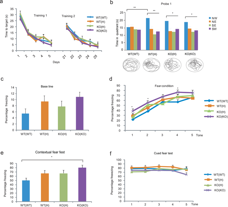
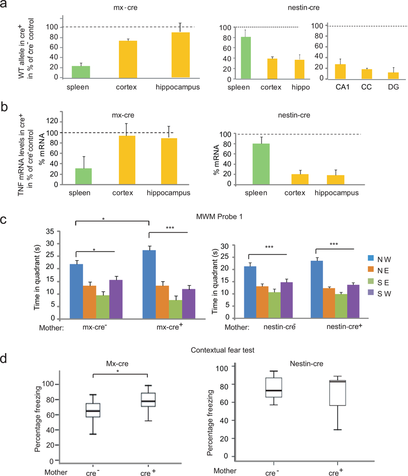
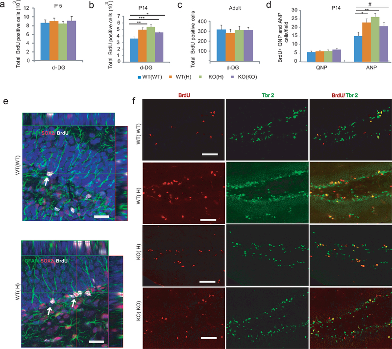
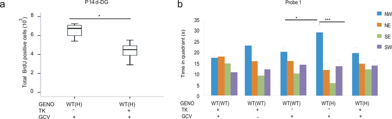
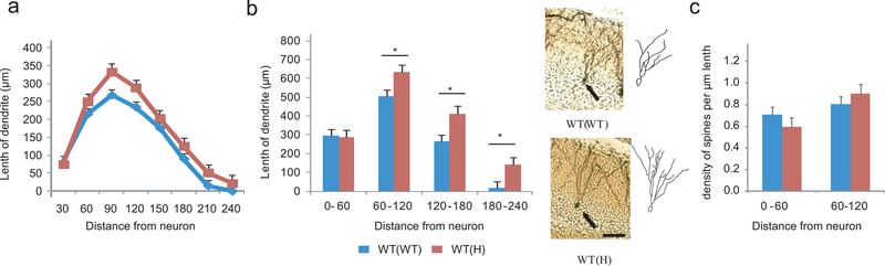
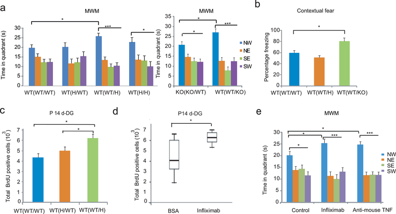
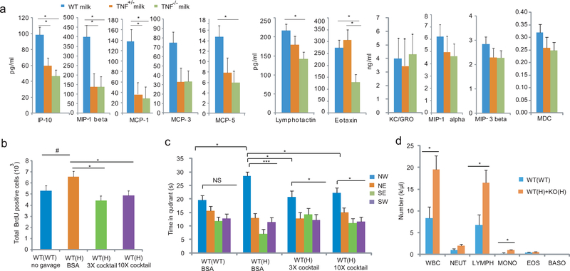
Tumor necrosis factor α (TNF) is a proinflammatory cytokine with established roles in host defense and immune system organogenesis. We studied TNF function and found a previously unidentified physiological function that extends its effect beyond the host into the developing offspring. A partial or complete maternal TNF deficit, specifically in hematopoietic cells, resulted in reduced milk levels of the chemokines IP-10, MCP-1, MCP-3, MCP-5 and MIP-1β, which in turn augmented offspring postnatal hippocampal proliferation, leading to improved adult spatial memory in mice. These effects were reproduced by the postpartum administration of a clinically used anti-TNF agent. Chemokines, fed to suckling pups of TNF-deficient mothers, restored both postnatal proliferation and spatial memory to normal levels. Our results identify a TNF-dependent 'lactrocrine' pathway that programs offspring hippocampal development and memory. The level of ambient TNF is known to be downregulated by physical activity, exercise and adaptive stress. We propose that the maternal TNF-milk chemokine pathway evolved to promote offspring adaptation to post-weaning environmental challenges and competition.


Conflict of interest statement

Competing Financial Interests

The authors declare no competing financial interests.

Figures

Figure 1.
Figure 1.
Maternal TNF deficit enhances cognitive functions in the offspring. (a) Training in the MWM with hidden platform. Although there was an effect of session in both the first and second training period (Repeated measures ANOVA: F4,752=73.38, P<10−5 and F4,640=45.29, P<10−5, respectively; N= 8,11,12,13/group), there was no genotype or genotype x session effect and consequently no significant difference between the groups at any time point. Data are from two independent experiments Data are shown as means ± SE. (b) In probe trial 1, two way ANOVA showed a significant effect of quadrant (F3,168=11.85, P<10−5) and LSD posthoc. test indicated that only the offspring of H and KO parents (but not those of WT parents) spent more time in the NW quadrant, the location that previously contained the platform (*p<0.05, **<0.005, ***<0.0005, NS=not significant; N= 8,11,12,13/group). To be considered as a significant change, time in NW target quadrant had to be significantly different from the time in all three other quadrants (lowest significance level among the three is displayed). The trace of movements is also displayed at the bottom of the panel. NW, North-West; NE, North-East; SE, South-East; SW, South-West. (c) Contextual and cued fear responses of the offspring of TNF H and KO parents. Baseline freezing (before conditioning) was not significantly different between the groups (ANOVA: F3,47=2.13, P=0.11; N=11,11,14,15/group). Data from three independent experiments (d) There was both a session- and a genotype-effect on freezing during context conditioning (Repeated measures ANOVA: F4.188=64.43, P<10−5 and F3.47=3.70, P=0.018, respectively) with KO(KO) offspring showing increased freezing compared to WT(WT) and WT(H) animals at the indicated time points (LSD posthoc; *p<0.05 vs. WT(WT) and #<0.05 vs. WT(H)). However, at the end of the training, the KO(KO) group was no longer different. (e) KO(KO) offspring showed an increase in contextual fear response (ANOVA: F3,47=4.25, P=0.010. Dunett T3 posthoc test; *p=0.006. When baseline measures were included as a covariate, the increase in contextual fear reaction in KO(KO) mice was still significant P=0.022). (f) The TNF deficient maternal environment has no effect on cued fear conditioning (F3,47:= 1.06, P=0.38).
Figure 2.
Figure 2.
Hematopoietic system specific inactivation of the TNF gene in the mother results in enhanced memory. (a) Inducible mx-cre expression results in recombination at the floxed TNF WT allele seen as a reduction in the overall level of the WT allele in the spleen but not in brain. In contrast, nestin-cre expression results in a substantial reduction of the WT allele in brain (e.g. cortex and hippocampus), in neurons (e.g. CA1 and DG of the hippocampus) and glia (e.g. corpus callosum=CC) but not in spleen. Hip=hippocampus. Data are shown as means ± SE. (b) Reduction of TNF mRNA is limited to the spleen in mx-cre+ animals while it is brain specific in nestin-cre+ mice. (c) Probe trial 1 of the MWM with the offspring of mx-cre+ and nestin-cre+ mothers. There was an effect of the platform location in both groups of animals (ANOVA: F3,56=35.69, P<10−5 and F3,80=42.43, P<10−5, respectively; N=7,9 and N=9,13 /group), but group x location interaction was seen only with the offspring of mx-cre+ mothers (F3,56=3.09, P=0.03) and LSD test showed that offspring of mx-cre+ as compared to the offspring of mx-cre- mothers had an increased memory of the platform location (*p<0.05;*** p< 0.0005). Data from three independent experiments (d) Increased freezing of the offspring of mx-cre+ (N=13,13/group) but not nestin-cre+ mothers (N=7,10/group) during contextual fear testing, t-test *p=0.014 and p=0.42 for mx-cre and nestin-cre, respectively. Box-whisker plots represent the first three quartiles (25%, median and 75%) and values 1.5× the interquartile range below the first quartile (lower horizontal line) and above the third quartile (upper horizontal line).
Figure 3.
Figure 3.
Increased proliferation in the developing DG is linked to enhanced adult spatial memory in the offspring of TNF mutant mothers. (a-c) Proliferation in the dorsal (d) subgranular zone (SGZ) at P5, P14 and adult as measured 2h after BrdU labeling. At P14, ANOVA showed a group difference in the number of BrdU positive cells (F3,16=7.38, P=0.003, N=5/group; LSD posthoc. *<0.05, **<0.005, ***<0.0005). Data are shown as means ± SE. (d) Maternal TNF genotype has no effect on QNP proliferation (F3,16=0.69, P=0.57) but increases ANP proliferation (F3,56=7.9, P<0.001; N=5/group; LSD posthoc. *p<0.05, **<0.005, #<0.1) at P14. (e) Confocal micrographs of WT(WT) and WT(H) SGZs with arrows showing QNPs characterized by BrdU and Sox-2 positivity and GFAP positive apical extensions toward the molecular layer of the DG. Bar=20μm. (f) Representative micrographs showing an increased number of BrdU+/Tbr2+ ANPs in the SGZ of WT(H), KO(H) and KO(KO) offspring as compared to the WT(WT) offspring at P14. Bar=50μm.
Figure 4.
Figure 4.
Genetic compensation of increased proliferation in the WT(H) offspring normalizes spatial memory. (a) GCV administered to neonates at P5 and P6 (12.5 mg/kg s.c.) resulted in an approximately 30% reduction in proliferation in P14 WT(H) offspring (F1,9=13.8, P=0.008; N=5,12/group). Box-whisker plots represent the first three quartiles (25%, median and 75%) and values 1.5× the interquartile range below the first quartile (lower horizontal line) and above the third quartile (upper horizontal line). Data are from two independent experiments. (b) In probe trial 1, control WT(WT) offspring with or without TK and in the presence or absence of GCV failed to learn the platform location. WT(H) offspring with no TK but injected with GCV could recall the platform location (two way ANOVA: quadrant F3,148=21.27, P<0.0001; group x quadrant, F12,148=1.9, P=0.04; N=6,7,8,10,11/group; LSD posthoc, *p<0.05, ***<0.0005). However, these offspring, if also harboring the TK gene, showed no learning. Data are shown as means ± SE.
Figure 5.
Figure 5.
Dendritic morphology-changes in adult WT(H) granule cells. (a) Dendritic length as a function of distance from the neuron in adult WT(WT) and WT(H) neurons. Two way ANOVA: group F1,288=10.94, P=0.0001. Neuron number=16,23 per genotype. Data are shown as means ± SE. (b) Increased dendritic length at distal areas in adult WT(H) granule cells. Two way ANOVA: group F1,148=13.42, P=0.001; LSD posthoc, *p<0.05. Representative images and corresponding tracings of a WT(WT) and a WT(H) neuron. Scale bar=20μm. (c) Spine density in adult WT(H) and WT(WT) granule cells. Two way ANOVA: group F1,66=0.13, P=0.91; distance from neuron F1,66=6.62, P=0.012. Neuron number=14,21 per genotype.
Figure 6.
Figure 6.
A. The maternal effect on offspring phenotypes is postnatal. (a,b) Association of the postnatal TNF deficient maternal environment with enhanced cognitive performance in MWM (Two way ANOVA; platform locationleft: F3,124=21.70, P<10−5, N= 12,6,12,5/group; platform locationright: F3,48=26.050, P=0.0001 and location x group F3,38=3.78, P=0.016, N=8,6; LSD posthoc. *p<0.05; ***<0.0005) and fear conditioning (ANOVA: F2, 21=4.32, P=0.03; LSD posthoc *p<0.05; N= 6,7,10). Data are shown as means ± SE. (c) Association of the postnatal TNF deficient maternal environment with increased proliferation in P14 DG (ANOVA: F2,16=6.3, P=0.01; N= 6,7,6; LSD posthoc. *p<0.05). (d) Postpartum administration of infliximab increases P14 DG proliferation in the offspring (t-test, T=2.709 p=0.014, N=14,6). Data are from two independent experiments. Box-whisker plots represent the first three quartiles (25%, median and 75%) and values 1.5× the interquartile range below the first quartile (lower horizontal line) and above the third quartile (upper horizontal line). (e) Offspring of infliximab and anti-mouse TNF antibody treated mothers had a higher level of recall of the platform location in probe trial 1 than that of the control offspring (Two way ANOVA; quadrant: F3,132=35.93, P<10−5; group x quadrant: F6,132=2.14, P=0.05; N=16,9,11/group; LSD posthoc. *p<0.05; ***p<0.0005). Controls, derived from mothers injected postnatally with either BSA or IgG1 isotype control antibodies, were pooled as their behavior did not differ.
Figure 7.
Figure 7.
Reduced milk chemokine levels in TNF deficient mothers are responsible for the WT(H) phenotypes. (a) Chemokine levels in postpartum day2 milk of TNF TNF+/− and TNF−/− mothers (two way ANOVA; genotype effect: F2,110=26.40, P<0.0001; group x chemokine: F20,110=4.31, P<0.0001; Tukey HSD posthoc test *p<0.05; N=5,3,5/group). Data are shown as means ± SE. (b) A cocktail of 5 recombinant cytokines at 3x and 10x doses given by gavage daily (1x: IP-10, 4 pg/g mouse weight; MIP-1β, 25pg/g; MCP-1, 10pg/g; MCP-3, 4 pg/g, and MCP-5, 0.7 pg/g) between P1 and P14 reduced proliferation in P14 DG (ANOVA; F3,118=4.14, P=0.021; N=5,6,6,5/group, LSD posthoc. *p<0.05, #<0.1). (c) The cytokine cocktails given between P1 and P21 reduced adult MWM memory in probe trial 1 at the 3x and 10x doses (Two way ANOVA; platform location: F3,75=24.98, P<10−5; group x platform location: F6,75=2.60, P=0.024; N=8,5,7,9/group, LSD posthoc. *p<0.05; ***<0.0005). (d) Increased WBC counts (t-test, T=2.678, p=0.019), due to elevated levels of lymphocyte and monocyte numbers in P10 pup of TNF+/− mothers as compared to pups of WT mothers (Manual count; Two way ANOVA, group: F1,65=9.37, P=0.0003; N=6,9/group, LSD posthoc. *p<0.05; **<0.005). Because the WT and KO pups of H mothers were not different, data were combined. NEUT, neutrophil; LYMPH, lymphocyte; MONO, monocyte; EOS, eosinophil; BASO, basophil.

Comment in

References

    1. Pasparakis M, Alexopoulou L, Episkopou V & Kollias G Immune and inflammatory responses in TNF alpha-deficient mice: a critical requirement for TNF alpha in the formation of primary B cell follicles, follicular dendritic cell networks and germinal centers, and in the maturation of the humoral immune response. The Journal of experimental medicine 184, 1397–1411 (1996). - PMC - PubMed
    1. Kuprash DV, et al. Novel tumor necrosis factor-knockout mice that lack Peyer’s patches. European journal of immunology 35, 1592–1600 (2005). - PubMed
    1. Muller N & Ackenheil M Psychoneuroimmunology and the cytokine action in the CNS: implications for psychiatric disorders. Prog Neuropsychopharmacol Biol Psychiatry 22, 1–33 (1998). - PubMed
    1. Stellwagen D & Malenka RC Synaptic scaling mediated by glial TNF-alpha. Nature 440, 1054–1059 (2006). - PubMed
    1. Golan H, Levav T, Mendelsohn A & Huleihel M Involvement of tumor necrosis factor alpha in hippocampal development and function. Cereb Cortex 14, 97–105 (2004). - PubMed

Publication types

MeSH terms

Associated data