. 2014 Mar;31(3):269-83.
doi: 10.1007/s10585-013-9626-1. Epub 2013 Dec 1.

Osteoblasts stimulate the osteogenic and metastatic progression of castration-resistant prostate cancer in a novel model for in vitro and in vivo studies

Affiliations

Osteoblasts stimulate the osteogenic and metastatic progression of castration-resistant prostate cancer in a novel model for in vitro and in vivo studies

Malin Hagberg Thulin et al. Clin Exp Metastasis. 2014 Mar.

Abstract

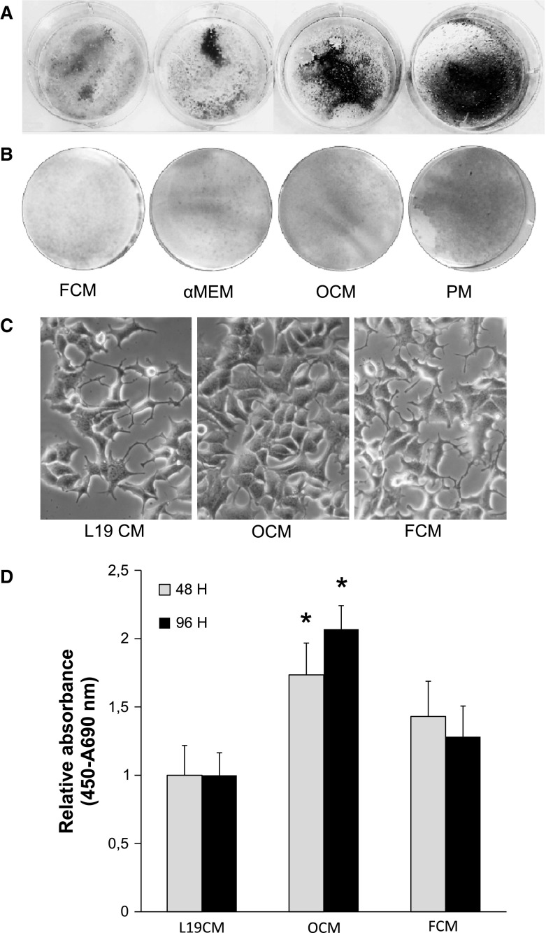
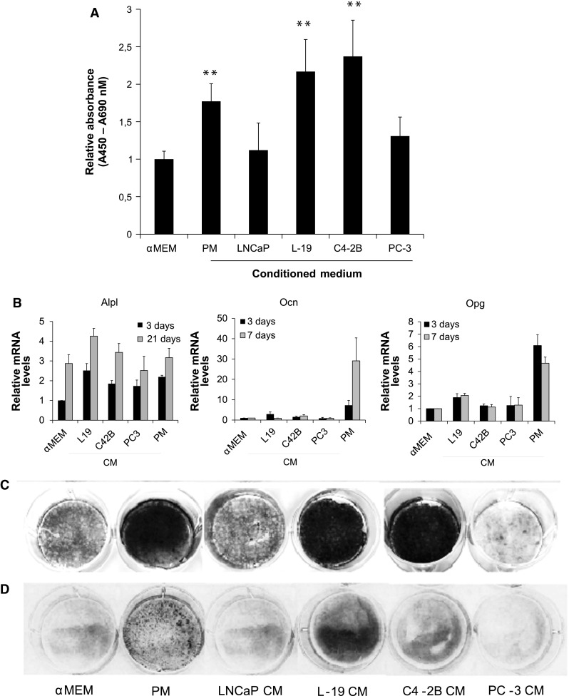
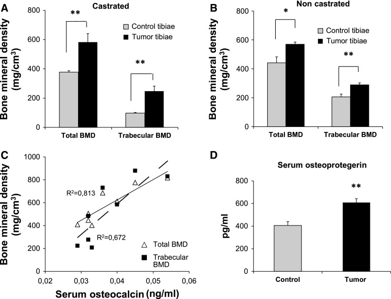
Castration-resistant prostate cancer (CRPC) is strongly associated with sclerotic bone metastases and poor prognosis. Models that mimic human CRPC are needed to identify the mechanisms for prostate cancer (PC) growth in bone and to develop new therapeutic strategies. We characterize a new model, LNCaP-19, and investigate the interaction between tumor cells and osteoblasts in the sclerotic tumor response of CRPC. Osteogenic profiling of PC cell lines (LNCaP-19, LNCaP, C4-2B4, and PC-3) was performed by gene expression arrays and mineral staining. Conditioned medium from MC3T3-E1 was used for osteoblast stimulation of CRPC cells. The capacity of LNCaP-19 cells to induce sclerotic lesions was assessed in intratibial xenografts and verified by serum markers, histological analysis and bone mineral density (BMD) measurements. The CRPC cell line LNCaP-19 expresses a pronounced osteogenic profile compared to its parental androgen-dependent cell line LNCaP. Osteoblast-derived factors further increase the expression of genes known to enhance metastatic progression of PC. LNCaP-19 forms sclerotic tumors in tibia of castrated mice as evident by increased total BMD (P < 0.01). There was a strong correlation between serum osteocalcin and BMD (total: R (2) 0.811, P < 0.01, trabecular: R (2) 0.673, P < 0.05). For the first time we demonstrate that a CRPC cell line generated in vitro has osteogenic capacity and that osteomimicry can be an inherent feature of these cells. Osteoblast-derived factors further promote the osteogenic and metastatic phenotype in CRPC cells. Altogether, our model demonstrates that both tumor cells and osteoblasts are mediators of the bone forming process of CRPC.


Figures

Fig. 1
Fig. 1
Osteoblasts induce LNCaP-19 mineralization and proliferation. LNCaP-19 cells were seeded in 6-well plates (1 × 10cells/well) and in 12-well plates (4 × 104 cells/well) for von Kossa staining respectively alkaline phosphatase (ALP) staining and cultured with fibroblast-conditioned medium (FCM), control medium (αMEM), osteoblast-conditioned (OCM), or promineralization medium (PM; αMEM supplemented with ascorbic acid (50 μg/ml) and β-glycerophosphate) (10 mM) under steroid deprived conditions (10 % FBS-DCC). a Mineralization of LNCaP-19, visualized after 21 days by von Kossa staining. b Staining of ALP activity after 21 days in culture. c Morphological changes in LNCaP-19 after 48 h in culture with OCM, FCM or control medium (L19CM) visualized with phase contrast light microscopy, magnification ×200. d Proliferation of LNCaP-19 cells. For proliferation, LNCaP-19 cells were seeded in 96-well plates (5 × 103 cells/well) and stimulated with control medium (L19 CM), OCM or FCM. All media was supplemented with 1 % FBS-DCC. Proliferation of LNCaP-19 was measured by BrdU incorporation after 48 and 96 h. Bars represent mean ± SEM of three independent experiments. *P < 0.05 versus control
Fig. 2
Fig. 2
LNCaP-19 cells affect osteoblast proliferation and mineralization. a Proliferation of MC3T3-E1 (3.5 × 103 cells per well in 96 well plates) was measured by BrdU incorporation after 5 days in culture with αMEM supplemented with 75 % CM. Bars represent mean ± SEM of three independent experiments. **P < 0.01 versus control medium (αMEM). Total RNA was extracted from MC3T3-E1 pre-osteoblasts at specified days and analyzed for the expression of differentiation markers by RT-qPCR. b Alp1 day 3, and 21, Ocn at day 3 and 7, and Opg at day 3 and 7. The expression levels were normalized to those in control medium (αMEM). Results are presented as mean ± SEM of three independent experiments. c For mineralization assays, MC3T3-E1 pre-osteoblasts were seeded in 6-well plates (1 × 105 cells/well) and in 12-well plates (4 × 104 cells/well), for von Kossa staining respectively alkaline phosphatase (Alp) staining. Cells were treated with control medium (αMEM), conditioned medium (CM) from PC cell lines, or promineralization medium (PM; αMEM supplemented with ascorbic acid (50 μg/ml) and β-glycerophosphate) (10 mM). All media were supplemented with 10 % FBS. Mineralization of MC3T3-E1 was visualized using von Kossa staining after 21 days in culture. d Staining of Alp activity detected after 21 days in culture
Fig. 3
Fig. 3
In vivo properties of LNCaP-19 evaluated as intratibial xenografts after 10 weeks of growth. LNCaP-19 cells (4 × 105) were inoculated into tibia of castrated and non-castrated nude BALB/c mice. The osteosclerotic tumor response was evaluated after 10 weeks. a, b pQCT measurements demonstrate increased total and trabecular BMD in tumor-bearing tibiae compared to control tibiae in a castrated (n = 9) and b non-castrated (n = 8) mice. c Mouse-specific ELISA shows correlation between serum osteocalcin (Ocn) concentrations and BMD in castrated mice, for total BMD (P < 0.01) and trabecular BMD (P < 0.05), while no correlation was detected in non-castrated mice (data not shown). d The osteoprotegerin (Opg) levels as a response to LNCaP-19 tumors in both castrated and non-castrated tumor bearing mice (n = 8 + 9) were measured by mouse-specific ELISA and compared to control mice. Bars represent mean ± SEM. **P < 0.002
Fig. 4
Fig. 4
Histology of the sclerotic tumor response of LNCaP-19. LNCaP-19 cells (4 × 105) were inoculated into tibia of castrated and non-castrated nude BALB/c mice. The osteosclerotic tumor response was evaluated after 10 weeks by histological evaluation of LNCaP-19 in tibiae of castrated and non-castrated mice. a, d Longitudinal sections (H&E staining) of LNCaP-19 in the tibia, demonstrating new formed bone replacing the bone marrow cavity (magnification ×200). b, e Control tibiae without tumor cell injection (magnification ×200). c, f Islands of interlaced LNCaP-19 cells within the newly formed trabecular bone (magnification ×400). ac represent tibiae from castrated mice and df non-castrated

References

    1. Michaelson MD, Marujo RM, Smith MR. Contribution of androgen deprivation therapy to elevated osteoclast activity in men with metastatic prostate cancer. Clin Cancer Res. 2004;10(8):2705–2708. doi: 10.1158/1078-0432.CCR-03-0735. - DOI - PubMed
    1. Vessella RL, Corey E. Targeting factors involved in bone remodeling as treatment strategies in prostate cancer bone metastasis. Clin Cancer Res. 2006;12(20 Pt 2):6285s–6290s. doi: 10.1158/1078-0432.CCR-06-0813. - DOI - PubMed
    1. Oefelein MG, Ricchuiti V, Conrad W, Seftel A, Bodner D, Goldman H, Resnick M. Skeletal fracture associated with androgen suppression induced osteoporosis: the clinical incidence and risk factors for patients with prostate cancer. J Urol. 2001;166(5):1724–1728. doi: 10.1016/S0022-5347(05)65661-3. - DOI - PubMed
    1. Mundy GR. Metastasis to bone: causes, consequences and therapeutic opportunities. Nat Rev Cancer. 2002;2(8):584–593. doi: 10.1038/nrc867. - DOI - PubMed
    1. Roodman GD. Mechanisms of bone metastasis. N Engl J Med. 2004;350(16):1655–1664. doi: 10.1056/NEJMra030831. - DOI - PubMed

Publication types

Substances

LinkOut - more resources