. 2014 Jan;11(1):106-12.
doi: 10.1038/nmeth.2737. Epub 2013 Dec 1.

Niche-independent high-purity cultures of Lgr5+ intestinal stem cells and their progeny

Affiliations

Niche-independent high-purity cultures of Lgr5+ intestinal stem cells and their progeny

Xiaolei Yin et al. Nat Methods. 2014 Jan.

Abstract

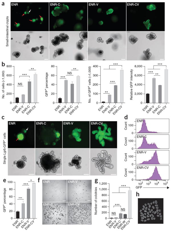
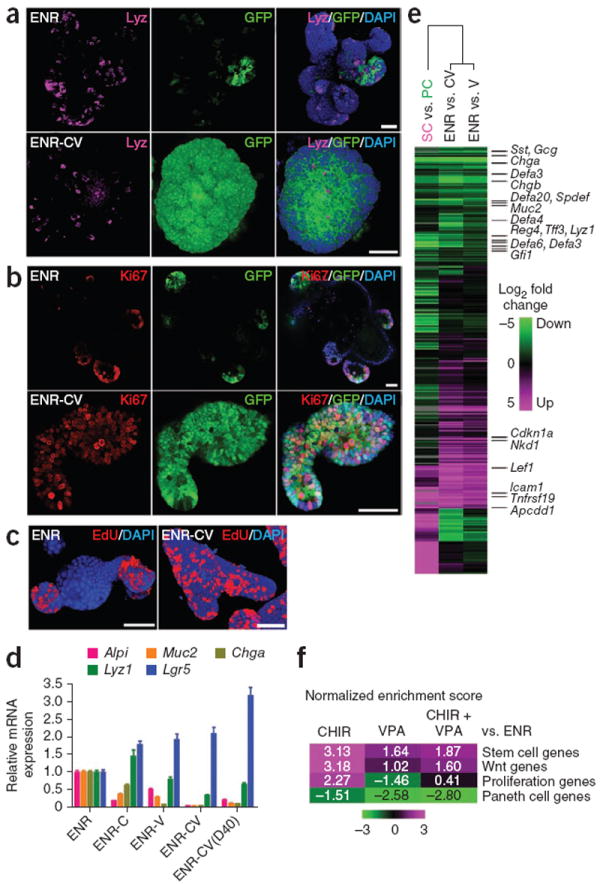
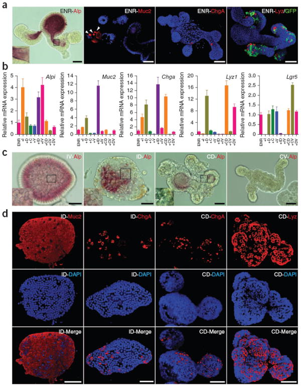
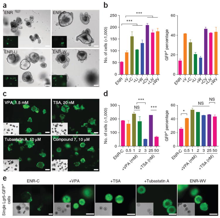
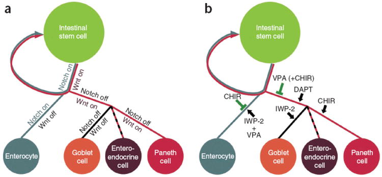
Although Lgr5(+) intestinal stem cells have been expanded in vitro as organoids, homogeneous culture of these cells has not been possible thus far. Here we show that two small molecules, CHIR99021 and valproic acid, synergistically maintain self-renewal of mouse Lgr5(+) intestinal stem cells, resulting in nearly homogeneous cultures. The colony-forming efficiency of cells from these cultures is ~100-fold greater than that of cells cultured in the absence of CHIR99021 and valproic acid, and multilineage differentiation ability is preserved. We made use of these homogeneous cultures to identify conditions employing simultaneous modulation of Wnt and Notch signaling to direct lineage differentiation into mature enterocytes, goblet cells and Paneth cells. Expansion in these culture conditions may be feasible for Lgr5(+) cells from the mouse stomach and colon and from the human small intestine. These methods provide new tools for the study and application of multiple intestinal epithelial cell types.


Conflict of interest statement

COMPETING FINANCIAL INTERESTS

The authors declare no competing financial interests.

Figures

Figure 1
Figure 1
The combination of CHIR and VPA promotes self-renewal of Lgr5+ stem cells. (a) GFP fluorescence and bright-field images of small-intestinal crypts cultured for 6 d in the presence of EGF, Noggin and R-spondin 1 (ENR); ENR and VPA (ENR-V); ENR and CHIR (ENR-C); and ENR, VPA and CHIR (ENR-CV). Apoptotic cells are visible in the lumen with autofluorescence (red arrows), and white arrow indicates Lgr5-GFP at the bottom of crypts. (b) Quantification of cell proliferation (number of live cells) and GFP expression in crypts cultured for 6 d. Lgr5-GFP expression was measured by flow cytometry. (c) Fluorescence and bright-field images of single Lgr5-GFP cells after 9 d of culture in multiple conditions. (d,e) Flow cytometry analysis of GFP expression 7 d after seeding of single Lgr5-GFP+ cells in conditions as indicated. Error bars, s.d. (n = 3 wells). (f,g) Representative images of 4,000 FACS-isolated single Lgr5-GFP cells cultured under one of several conditions for 7 d (f) and quantification of colony numbers (g). (h) Metaphase spread of a cell cultured in the CV condition for 80 d. Scale bars, 100 μm (a,c) and 1 mm (f). ***P < 0.001; **P < 0.01; * P < 0.05; NS, not significant (P > 0.05). Error bars, s.d. (n = 3 wells). Experiments in b and e were performed in three biological replicates (n = 3 animals), with similar results (data not shown).
Figure 2
Figure 2
The combination of CHIR and VPA maintains the stem cell state of Lgr5+ stem cells. (a–c) Confocal images showing lysozyme (a), Ki67 (b) and EdU (c) staining of organoids cultured under the ENR and ENR-CV conditions. DAPI was used as a nuclear stain. (d) Quantitative real-time PCR analysis of relative mRNA expression of markers for intestinal epithelial cells (Alpi for enterocytes, Muc2 for goblet cells, Chga for enteroendocrine cells, Lyz1 for Paneth cells and Lgr5 for ISCs) cultured for 6 d under conditions as indicated. ENR-CV(D40) indicates cells that were cultured in the CV condition for 40 d. Error bars, s.d. (n = 3 wells). Experiments were performed in three biological replicates (n = 3 animals), with similar results (data not shown). (e) Microarray analysis of gene expression changes in organoids cultured for 6 d in the presence of ENR-V (V) or ENR-CV (CV) compared with ENR. The heat map shows clustering to previously reported array data of sorted Paneth cells (PC) versus Lgr5 stem cells (SC). Green and magenta indicate down- and upregulation, respectively. (f) Gene-set enrichment analysis (GSEA) comparing organoid microarray data with published gene expression signatures. Normalized enrichment scores are represented as a heat map to visualize the degree of enrichment in the up- or downregulated genes in organoids (magenta and green, respectively). Refer to Supplementary Figure 5 for detailed analysis. Scale bars, 50 μm.
Figure 3
Figure 3
Differentiation of ISCs cultured under the CV condition. (a) Staining of the indicated differentiation markers of Lgr5-GFP cells transferred from the CV condition to ENR and cultured for 4 d. DAPI (blue) was used to stain nuclei, and GFP indicates the presence of stem cells. (b) Real-time reverse-transcription PCR analysis of relative mRNA expression of mature intestinal epithelial markers from cells initially cultured in CV (6 d) and then transferred to the indicated conditions. ENR was present in all conditions and mRNA expression was normalized to expression in ENR alone. I, IWP-2; D, DAPT; C, CHIR; V, VPA. Error bars, s.d. (n = 3 wells). (c) Alp staining of cells cultured under multiple conditions. The morphological change of cells in the ID and CD conditions reflects formation of goblet and Paneth cells, respectively. (d) Immunocytochemistry staining of differentiation markers. Cells cultured under the ID and CD conditions were used for Muc2, ChgA and Lyz staining. Three-dimensional reconstructed confocal images are shown. Scale bars, 50 μm.
Figure 4
Figure 4
Exploring the mechanism of action for CHIR and VPA. (a) Morphology and Lgr5-GFP expression of crypts cultured in multiple conditions for 6 d. C, CHIR; Li, LiCl; W, Wnt3a; V, VPA. (b) Cell numbers and percentage of GFP+ cells for 6-d crypt cultures. The data are representative of three independent experiments. Error bars, s.d. (n = 3 wells). (c) 6-d cultures of crypts in the ENR-C (control) condition or with histone deacetylase (HDAC) inhibitors. See Supplementary Figure 6 for complete analysis. TSA, trichostatin A. (d) Effects of VPA and TSA on cell proliferation and GFP expression at multiple concentrations. Error bars, s.d. (n = 2 wells). Experiments were performed in three biological replicates (n = 3 animals), with similar results (data not shown). (e) Effects of other HDAC inhibitors and recombinant Wnt protein. Morphology and GFP expression of single Lgr5-GFP cells cultured for 6 d are shown. TSA concentration is 25 nM; tubastatin A is 10 μM. Scale bars, 100 μm. ***P < 0.001; *P < 0.05; NS, not significant (P > 0.05).
Figure 5
Figure 5
Model for ISC self-renewal and differentiation. (a) Under physiological conditions, the self-renewal and differentiation of ISCs is controlled by the cooperation of Wnt and Notch pathways. (b) When Lgr5+ stem cells are cultured in vitro, CHIR activates the Wnt pathway and inhibits enterocyte differentiation, whereas VPA alone or together with CHIR suppresses secretory cell specification. The combination of CHIR and VPA maintains ISCs in an undifferentiated, self-renewing state. Additional combinations of inhibitors promote various intestinal cell fates as shown.

References

    1. Barker N, et al. Identification of stem cells in small intestine and colon by marker gene Lgr5. Nature. 2007;449:1003–1007. - PubMed
    1. Sato T, et al. Single Lgr5 stem cells build crypt-villus structures in vitro without a mesenchymal niche. Nature. 2009;459:262–265. - PubMed
    1. Sato T, et al. Paneth cells constitute the niche for Lgr5 stem cells in intestinal crypts. Nature. 2011;469:415–418. - PMC - PubMed
    1. Yilmaz ÖH, et al. mTORC1 in the Paneth cell niche couples intestinal stem-cell function to calorie intake. Nature. 2012;486:490–495. - PMC - PubMed
    1. Farin HF, van Es JH, Clevers H. Redundant sources of Wnt regulate intestinal stem cells and promote formation of Paneth cells. Gastroenterology. 2012;143:1518–1529. - PubMed

Publication types

MeSH terms

Associated data