Review
. 2013 Dec;57(1-3):268-78.
doi: 10.1007/s12026-013-8469-9.

Killing machines: three pore-forming proteins of the immune system

Affiliations
Review

Killing machines: three pore-forming proteins of the immune system

Ryan McCormack et al. Immunol Res. 2013 Dec.

Abstract

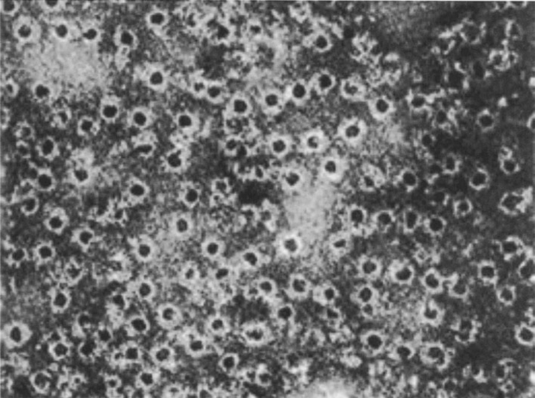
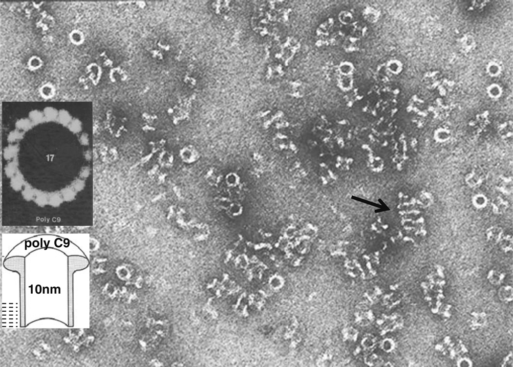
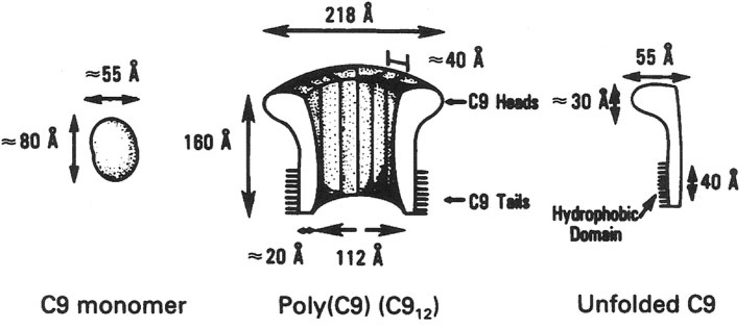
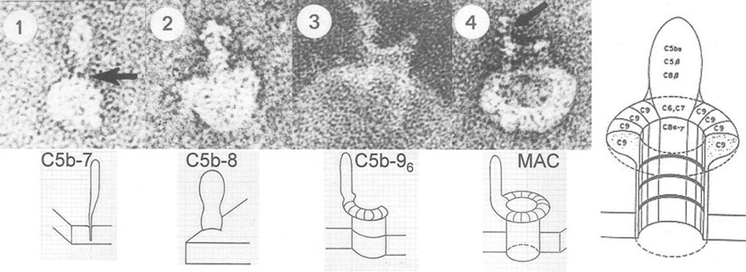
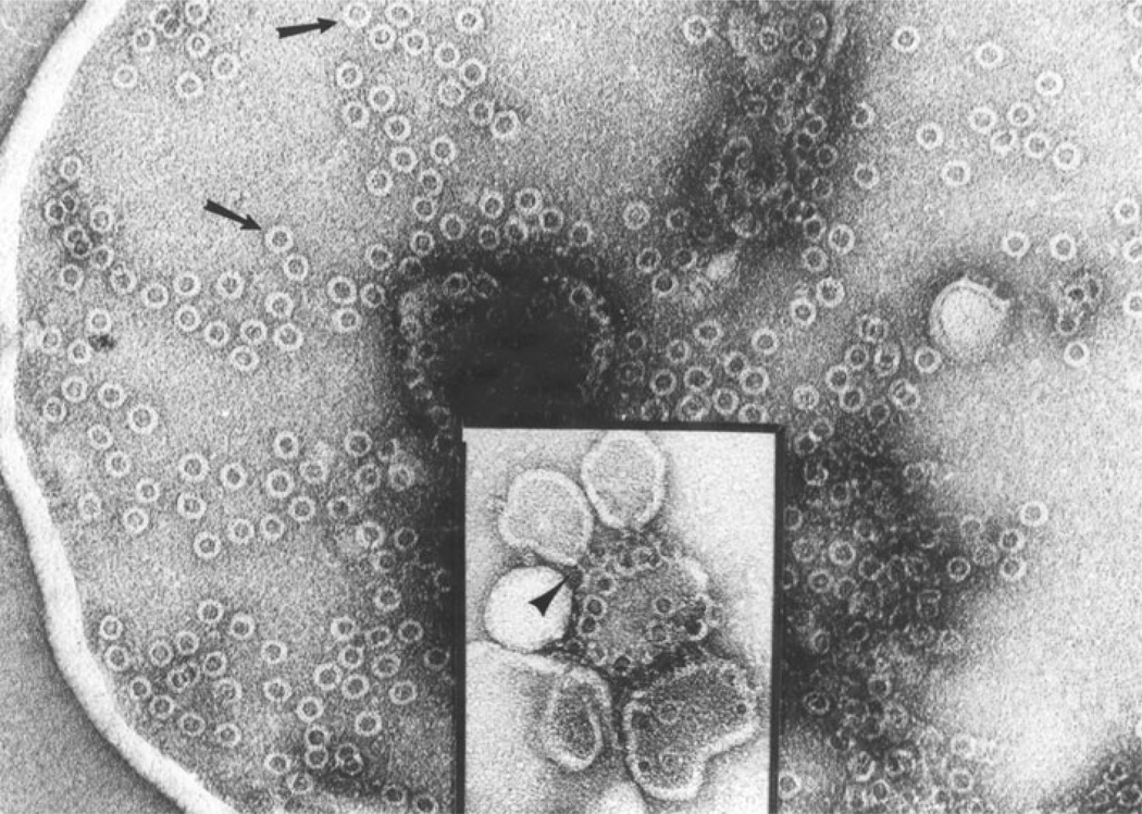
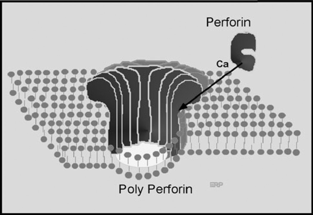
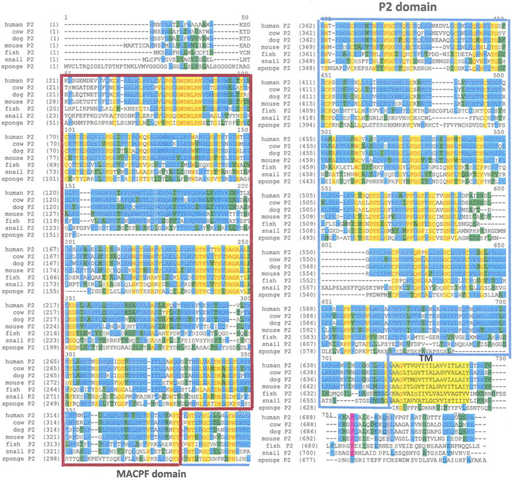
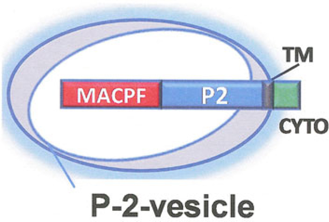
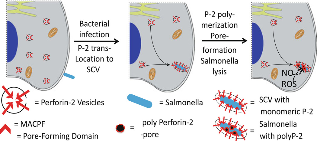
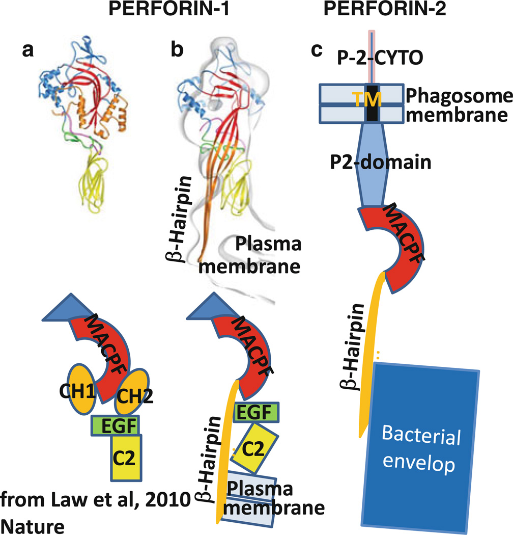
The evolution of early multicellular eukaryotes 400-500 million years ago required a defensive strategy against microbial invasion. Pore-forming proteins containing the membrane-attack-complex-perforin (MACPF) domain were selected as the most efficient means to destroy bacteria or virally infected cells. The mechanism of pore formation by the MACPF domain is distinctive in that pore formation is purely physical and unspecific. The MACPF domain polymerizes, refolds, and inserts itself into bilayer membranes or bacterial outer cell walls. The displacement of surface lipid/carbohydrate molecules by the polymerizing MACPF domain creates clusters of large, water-filled holes that destabilize the barrier function and provide access for additional anti-bacterial or anti-viral effectors to sensitive sites that complete the destruction of the invader via enzymatic or chemical attack. The highly efficient mechanism of anti-microbial defense by a combined physical and chemical strategy using pore-forming MACPF-proteins has been retargeted during evolution of vertebrates and mammals for three purposes: (1) to kill extracellular bacteria C9/polyC9 evolved in conjunction with complement, (2) to kill virus infected and cancer cells perforin-1/polyperforin-1 CTL evolved targeted by NK and CTL, and (3) to kill intracellular bacteria transmembrane perforin-2/putative polyperforin-2 evolved targeted by phagocytic and nonphagocytic cells. Our laboratory has been involved in the discovery and description of each of the three pore-formers that will be reviewed here.


Figures

Fig. 1
Fig. 1
Complement lesions on erythrocyte membranes as originally described by Humphrey et al. in 1964
Fig. 2
Fig. 2
Jürg Tschopp
Fig. 3
Fig. 3
PolyC9 in solution. The cylindrical complexes are seen in top view as white rings and in side views as hollow, negativestain-filled cylinders. The arrow points to an array of polymers that interact via the hydrophobic lipid-binding domains allowing the determination of the length (~50 Å) of the hydrophobicity area. Inset: heptadecameric polyC9, image-enhanced by rotational averaging
Fig. 4
Fig. 4
Model of monomeric and polymerized C9 with approximate dimensions and hydrophobic area, as published by us in 1982
Fig. 5
Fig. 5
Images of the growing membrane-attack complex of complement inserted into a single-layer lipid vesicle. Arrow in one points to the knife like lipid-binding domain of C5b–7. The arrow in four points to the C5b–8 part of MAC-polyC9
Fig. 6
Fig. 6
Gunther Dennert
Fig. 7
Fig. 7
Membrane lesions on membranes of cells killed by NK cells or CTL as published in 1983. The arrows show the cylindrical polyperforin-1 complex in top view, the arrow head in the inset in side view. The inner diameter is 160 Å
Fig. 8
Fig. 8
Model of perforin-1 polymerization and insertion into lipid bilayer triggered by extracellular calcium ions
Fig. 9
Fig. 9
Left: M. smegmatis from fresh liquid culture. Right: M. smegmatis isolated from IFN-activated fibroblasts 5 h after infection and then plated on agar overnight. Note the intact but grotesquely swollen body of M. smegmatis. Addition of lysozyme does not affect the images on the left but disintegrates M. smegmatis on the right to small fragments and debris (not shown)
Fig. 10
Fig. 10
Phylogenetic conservation of perforin-2 from sponge to man. Identity is indicated by red/yellow, conservative substitution is shown in blue and green The transmembrane domain is indicated as TM followed by the short cytoplasmic domain (Color figure online)
Fig. 11
Fig. 11
Predicted transmembrane orientation of perforin-2 in membrane vesicles stored in the cytoplasm of perforin-2 expressing cells
Fig. 12
Fig. 12
Hypothetical model of perforin-2 vesicles trafficking to and fusing with the bacterium-containing endosome/phagosome, perforin-2-polymerization and killing the bacterium by pore formation
Fig. 13
Fig. 13
Model of perforin-2 refolding and killing bacterium based on crystal structure of perforin-1 (Law et al. 2010). a Crystal structure and schematic domain structure of perforin-1. b Refolded perforin-1 inserted into lipid bilayer (plasma membrane). c Refolded perforin-2 attacking bacteria inside phagosome. Note that perforin-2 remains tethered to the phagosome membrane via its transmembrane domain

References

    1. Borsos T, Dourmashkin RR, Humphrey JH. Lesions in erythrocyte membranes caused by immune haemolysis. Nature. 1964;202:251–252. - PubMed
    1. Humphrey JH, Dourmashkin RR. The lesions in cell membranes caused by complement. Adv Immunol. 1969;11:75–115. - PubMed
    1. Mayer MM. Mechanism of cytolysis by complement. Proc Natl Acad Sci USA. 1972;69:2954–2958. - PMC - PubMed
    1. Bhakdi S, Tranum-Jensen J. Molecular nature of the complement lesion. Proc Natl Acad Sci USA. 1978;75:5655–5659. - PMC - PubMed
    1. Zalman LS, Wood LM, Muller-Eberhard HJ. Isolation of a human erythrocyte membrane protein capable of inhibiting expression of homologous complement transmembrane channels. Proc Natl Acad Sci USA. 1986;83:6975–6979. - PMC - PubMed

MeSH terms

Substances

LinkOut - more resources