. 2013 Nov;36(5):400-9.
doi: 10.1007/s10059-013-0078-x. Epub 2013 Nov 14.

(S)-tetrahydroisoquinoline alkaloid inhibits LPS-induced arachidonic acid release through downregulation of cPLA2 expression

Affiliations

(S)-tetrahydroisoquinoline alkaloid inhibits LPS-induced arachidonic acid release through downregulation of cPLA2 expression

Jong Min Choi et al. Mol Cells. 2013 Nov.

Abstract

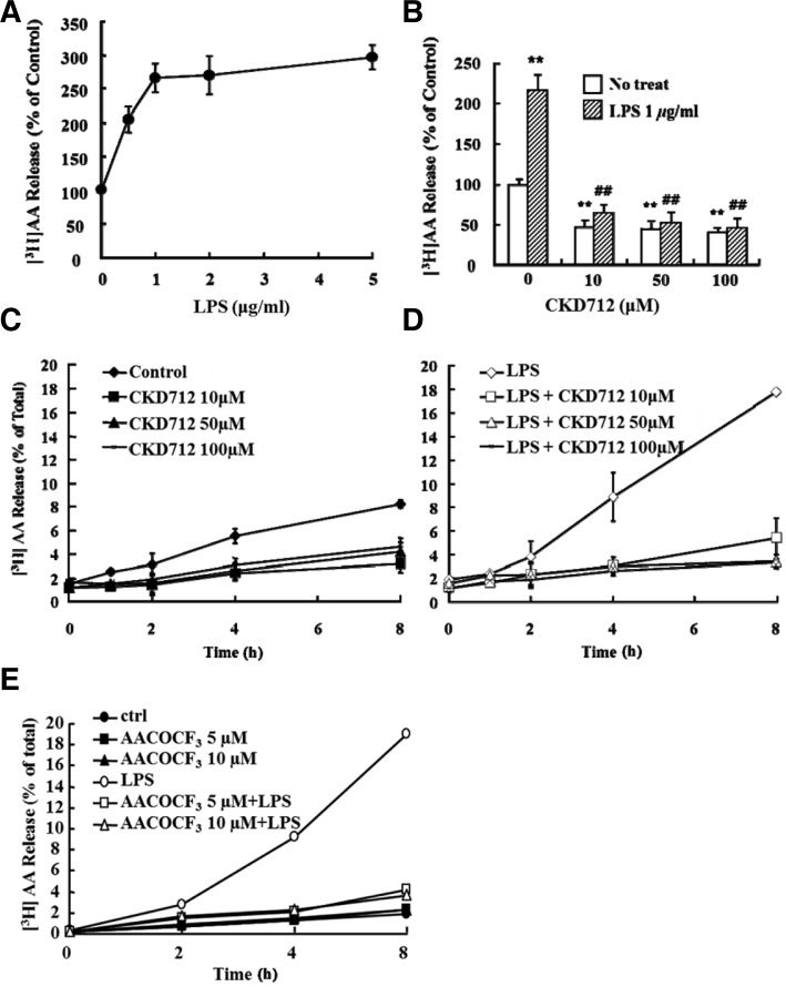
Sepsis, a systemic inflammatory response syndrome, remains a potentially lethal condition. (S)-1-α-Naphthylmethyl-6,7-dihydroxy-1,2,3,4-tetrahydroisoquinoline (CKD712) is noted as a drug candidate for sepsis. Many studies have demonstrated its significant anti-inflammatory effects. Here we first examined whether CKD712 inhibits lipopolysaccharide (LPS)-induced arachidonic acid (AA) release in the RAW 264.7 mouse monocyte cell line, and subsequently, its inhibitory mechanisms. CKD712 reversed LPS-associated morphological changes in the RAW 264.7 cells, and inhibited LPS-induced release of AA in a concentrationdependent manner. The inhibition was apparently due to the diminished expression of a cytosolic form of phospholipase A2 (cPLA2) by CKD712, resulting from reduced NF-κB activation. Furthermore, CKD712 inhibited the activation of ERK1/2 and SAP/JNK, but not of p38 MAPK. CKD712 had no effect on the activity or phosphorylation of cPLA2 and on calcium influx. Our results collectively suggest that CKD712 inhibits LPS-induced AA release through the inhibition of a MAPKs/NF-κB pathway leading to reduced cPLA2 expression in RAW 264.7 cells.


Figures

Fig. 1
Fig. 1
Structure of CKD712. (S)-1-α-Naphthylmethyl-6,7-dihydroxy-1,2,3,4-tetrahydroisoquinoline.
Fig. 2
Fig. 2
Effect of CKD712 on LPS-induced morphological changes in RAW 264.7 cells. RAW 264.7 cells were incubated for 1 h in serum-free DMEM, and then incubated with the indicated concentrations of CKD712. After 30 min, the cells were incubated with 1 μg/ml LPS for 2 h. Black arrows represent morphologically altered RAW 264.7 cells. The morphology of RAW 264.7 cells was visualized using optical microscopy (200× magnification).
Fig. 3
Fig. 3
Effect of CKD712 on LPS-induced [3H] AA release in RAW 264.7 cells. (A) RAW 264.7 cells were incubated for 24 h in DMEM containing 10% FBS with 0.5 μCi/ml [3H] AA, washed 3 times with free DMEM, and then incubated for 8 h with various concentrations of LPS. The supernatants were then collected and tested for radioactivity (Tri-Carb 2910TR/ Perkin Elmer LAS). The radioactivity released by untreated cells was considered 100%. (B) [3H] AA release induced by LPS in RAW 264.7 cells. RAW 264.7 cells were incubated for 24 h in complete DMEM with 0.5 μCi/ml [3H] AA, washed 3 times with free DMEM, and then incubated with the indicated concentrations of CKD712. After 1 h, incubated cells were treated with 1 μg/ml LPS and incubated for 8 h. The supernatants were then collected and the radioactivity was counted (Tri-Carb 2910TR/ Perkin Elmer LAS). The radioactivity released by untreated cells was considered 100%. After treatment with only CKD712 (C) or CKD712 and LPS (D), the [3H] AA released was detected in the supernatant over an 8-h time course (0, 1, 2, 4, and 8 h). (E) After treatment of AACOCF3 (0, 5, and 10 μM) with or without LPS treatment, released [3H] AA was detected in the supernatant over an 8-h time course (0, 2, 4, and 8 h). **p < 0.01 compared with no treated-control group, ##p < 0.01 compared with LPS and 0 μM-treated group.
Fig. 4
Fig. 4
CKD712 has no direct effect on purified cPLA2 from RAW 264.7 cells. cPLA2 was partially purified from RAW 264.7 cells following the procedure outlined in the “Materials and Methods”. (A) The prepared sample was applied to HiTrap TM Heparin HP column (1 ml, GE Healthcare) pre-equilibrated with buffer H at a flow rate of 0.5 ml/min. (B) The PT of Hi-Trap™ Heparin HP column, enzyme activity was recovered in unbound path-through (PT), was applied to Mono Q TM column (GE healthcare). (C) Purified cPLA2 was incubated with AACOCF3 (cPLA2 inhibitor) or DTT (sPLA2 inhibitor) or CKD712 at various concentration for 10 min, and then, PLA2 activity was assayed with the standard assay system.
Fig. 5
Fig. 5
CKD712 downregulates the activity and expression level but not phosphorylation of cPLA2. (A) RAW 264.7 cells were incubated for 1 h with the indicated concentrations of CKD712. After 1 h, the cells were treated with 1 μg/ml LPS and incubated for 8 h. Cells were then harvested and washed 2 times with TBS, and sonicated in lysis buffer (120 mM NaCl, 7 mM Tris-Cl, and 1 mM EDTA, pH 9.0). The sonicated cells were centrifuged at 2,000 × g for 10 min at 4°C. The supernatants (lysates) were assayed for cPLA2 activity, and immunobloted with anti-cPLA2 antibody. (B) RAW 264.7 cells were incubated with LPS for 30 min after pre-incubation with CKD712 at 0, 1, 10, 50, and 100 μM for 30 min. The lysate was then immunobloted with anti-cPLA2 and anti-phospho-cPLA2 antibodies. *p < 0.05, **p < 0.01 compared with no treated-control group, ##p < 0.01 compared with LPS and 0 μM-treated group.
Fig. 6
Fig. 6
Effect of CKD712 on cPLA2 activation by calcium influx. RAW 264.7 cells were plated in 96-well black plates at 5 × 10⁴ cells per well. After 24 h, calcium influx was detected with Fluo-4 NW Calcium Assay Kits (Invitrogen, USA) at 516 nm wavelength. Calcium influx into the cytosol was detected in cells treated with LPS (A) and A23187 (B) for 10 s (arrows).
Fig. 7
Fig. 7
Inhibition of LPS induced NF-κB activation by CKD712. After CKD712 was treated at indicated concentrations for 1 h in free DMEM, RAW 264.7 cells were treated with 1 μg/ml LPS and incubated for 4 h. Lysates and nuclear extracts of cells were prepared using a Nuclear Extraction kit (Panomics, USA). (A) Five micrograms of nuclear extract was incubated with a biotin-labeled probe; NF-κB: (5′ → 3′) AGT TGA GGG GAC TTT CCC AGG C, at 15°C for 30 min. Images of probes bound with nuclear extract were acquired using ChemiDoc™ XRS (Bio-Rad), and the lysates were immunobloted with anti-cPLA2 and anti-COX-2 antibodies (B) After incubation with CKD712 and LPS for 8 h, released PGE2 was detected, in the medium used to grow the cells, by using anti-PGE2 antibody (PGE2 ELISA kit; Sapphire Bioscience). **p < 0.01 compared with no treated-control group, ##p < 0.01 compared with LPS and 0 μM-treated group.
Fig. 8
Fig. 8
Inhibition of phosphorylation of ERK1/2 and JNK/SAP by CKD712. After the cells were treated with MAPK inhibitors at 5 μM (A) or CKD712 at 0, 1, 10, 50, and 100 μM (B) for 0.5 h in free DMEM, RAW 264.7 cells were treated with 1 μg/ml LPS and incubated for 0.5 h. Next, the cells were harvested and washed 2 times in TBS, and then sonicated in lysis buffer (120 mM NaCl, 75 mM Tris-Cl, and 1 mM EDTA, pH 9.0). The sonicated cells were centrifuged at 2,000 × g for 10 min at 4°C. Supernatants (lysates) were immunobloted with anti-MAPK, anti-phospho-MAPK, anti-cPLA2, and anti-phospho-cPLA2 antibodies. GAPDH was used as a protein-loading control; its phospho-protein form (A) was detected by anti-GAPDH, not by anti-phospho-GAPDH. For immunob-lotting of phospho-proteins, BSA was used instead of skim milk, and NaN3 was excluded from all process-sing.
Fig. 9
Fig. 9
LPS-induced AA release and cPLA2 expression is related to the MAPK-activating pathway. (A) [3H] AA release was induced by LPS in RAW 264.7 cells, which were incubated for 24 h in complete DMEM with 0.5 μCi/ml [3H] AA, washed 3 times with free DMEM, and then incubated with MAPK inhibitors or CKD712. MAPK inhibitors were added at IC50 concentrations that downregulate cytokine levels in monocytes: SP600125 (JNK/SAP inhibitor), IC50: 5–10 μM, SB203580 (p38 MAPK inhibitor), IC50: 50–100 nM, and PD98059 (ERK1/2 inhibitor), IC50: 2 μM. After 0.5 h, the incubated cells were treated with 1 μg/ml LPS and incubated for 8 h. [3H] AA was detected in the supernatant over an 8-h time course (0, 2, 4, 40, and 8 h), using Tri-Carb 2910TR/ Perkin Elmer LAS. (B) RAW 264.7 cells were incubated for 0.5 h with CKD712 or MAPK inhibitors. After 0.5 h, cells were treated with 1 μg/ml LPS and incubated for 8 h. Cells were then harvested and washed 2 times with TBS, followed by sonication in lysis buffer (12 mM NaCl, 75 mM Tris-Cl, and 1 mM EDTA, pH 9.0). The sonicated cells were centrifuged at 2,000 × g for 10 min at 4°C. The RAW 264.7 cell lysate was immunobloted with anti-cPLA2 antibody, and assayed for cPLA2 activity. **p < 0.01 compared with no treated-control group, #p < 0.05, ##p < 0.01 compared with LPS only-treated group.

References

    1. Barancik M, Bohacova V, Kvackajova J, Hudecova S, Krizanova O, Breier A. SB203580, a specific inhibitor of p38-MAPK pathway, is a new reversal agent of P-glycoprotein-mediated multidrug resistance. Eur J Pharm Sci. 2001;14:29–36. - PubMed
    1. Baron RM, Baron MJ, Perrella MA. Pathobiology of sepsis: are we still asking the same questions? Am J Respir Cell Mol Biol. 2006;34:129–134. - PubMed
    1. Bennett BL, Sasaki DT, Murray BW, O’Leary EC, Sakata ST, Xu W, Leisten JC, Motiwala A, Pierce S, Satoh Y, et al. SP600125, an anthrapyrazolone inhibitor of Jun N-terminal kinase. Proc. Natl. Acad. Sci. USA. 2001;98:13681–13686. - PMC - PubMed
    1. Casas J, Meana C, Esquinas E, Valdearcos M, Pindado J, Balsinde J, Balboa MA. Requirement of JNK-mediated phosphorylation for translocation of group IVA phospholipase A2 to phagosomes in human macrophages. J Immunol. 2009;183:2767–2774. - PubMed
    1. Clark JD, Schievella AR, Nalefski EA, Lin LL. Cytosolic phospholipase A2. J Lipid Mediat Cell Signal. 1995;12:83–117. - PubMed

Publication types