

Crystal structure of the tetrameric fibrinogen-like recognition domain of fibrinogen C domain containing 1 (FIBCD1) protein
- PMID: 24293368
- PMCID: PMC3908420
- DOI: 10.1074/jbc.M113.520577
Crystal structure of the tetrameric fibrinogen-like recognition domain of fibrinogen C domain containing 1 (FIBCD1) protein
Abstract
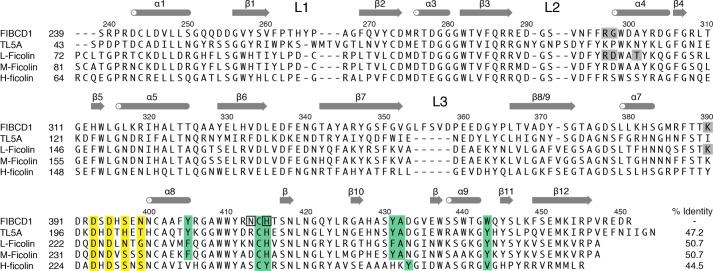
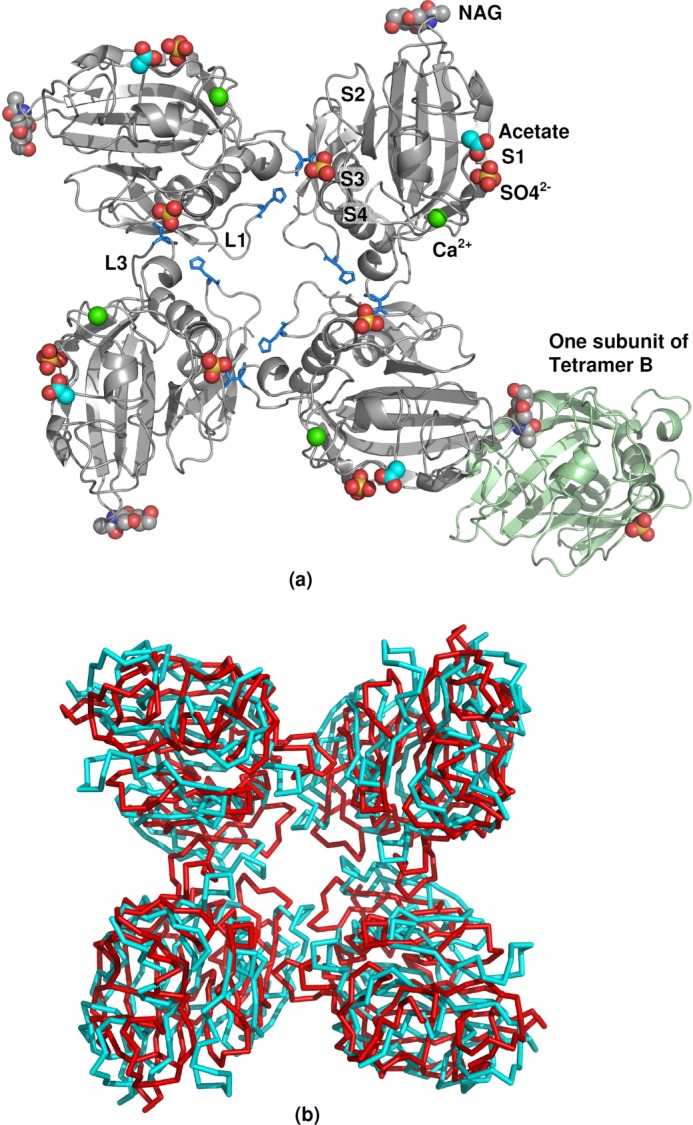
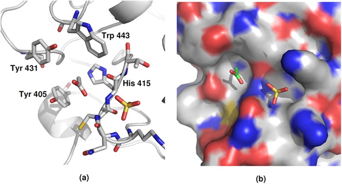
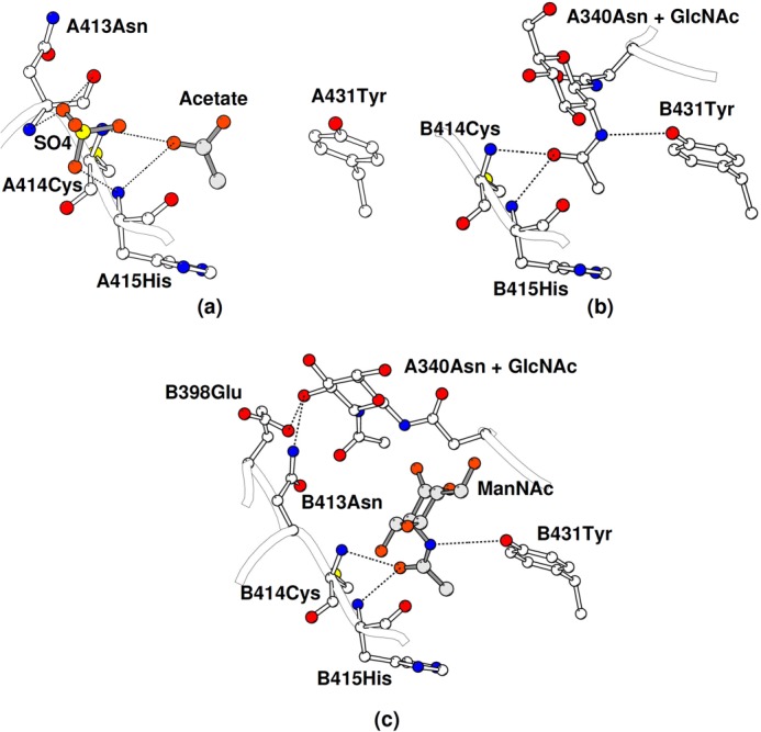
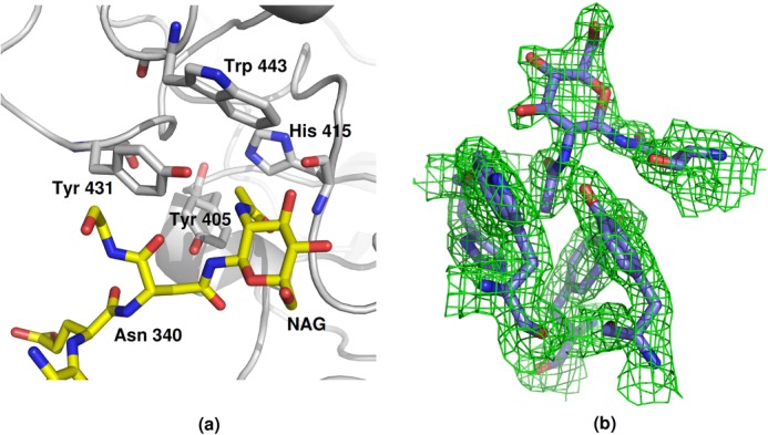
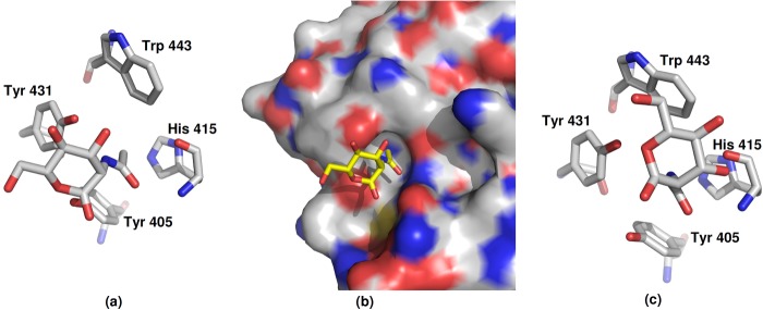
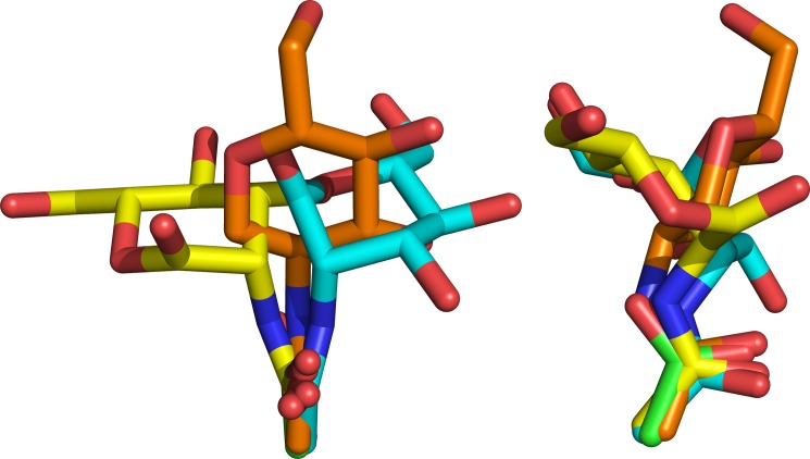
The high resolution crystal structures of a recombinant fragment of the C-terminal fibrinogen-like recognition domain of FIBCD1, a vertebrate receptor that binds chitin, have been determined. The overall tetrameric structure shows similarity in structure and aggregation to the horseshoe crab innate immune protein tachylectin 5A. The high affinity ligand N-acetylmannosamine (ManNAc) binds in the S1 site, predominantly via the acetyl group with the oxygen and acetamide nitrogen hydrogen-bonded to the protein and the methyl group inserted into a hydrophobic pocket. The binding of the ManNAc pyranose ring differs markedly between the two independent subunits, but in all structures the binding of the N-acetyl group is conserved. In the native structure, a crystal contact results in one of the independent protomers binding the first GlcNAc of the Asn(340) N-linked glycan on the other independent protomer. In the ligand-bound structure this GlcNAc is replaced by the higher affinity ligand ManNAc. In addition, a sulfate ion has been modeled into the electron density at a location similar to the S3 binding site in L-ficolin, whereas in the native structure an acetate ion has been placed in the S1 N-acetyl binding site, and a sulfate ion has been placed adjacent to this site. These ion binding sites are ideally placed to receive the N-acetyl and sulfate groups of sulfated GalNAc residues of glycosaminoglycans such as chondroitin and dermatan sulfate. Together, these structures give insight into important determinants of ligand selectivity, demonstrating versatility in recognition and binding while maintaining conservation in N-acetyl and calcium binding.
Keywords: Acetyl Binding; Chitin Receptor; Crystal Structure; FIBCD1; Fibrinogen-like Domain; Ligand-binding Protein; Pattern Recognition Receptor; Receptor Structure-Function; Structural Biology.
Figures
References
-
- Schlosser A., Thomsen T., Moeller J. B., Nielsen O., Tornøe I., Mollenhauer J., Moestrup S. K., Holmskov U. (2009) Characterization of FIBCD1 as an acetyl group-binding receptor that binds chitin. J. Immunol. 183, 3800–3809 - PubMed
-
- Thomsen T., Moeller J. B., Schlosser A., Sorensen G. L., Moestrup S. K., Palaniyar N., Wallis R., Mollenhauer J., Holmskov U. (2010) The recognition unit of FIBCD1 organizes into a noncovalently linked tetrameric structure and uses a hydrophobic funnel (S1) for acetyl group recognition. J. Biol. Chem. 285, 1229–1238 - PMC - PubMed
-
- Krarup A., Thiel S., Hansen A., Fujita T., Jensenius J. C. (2004) L-ficolin is a pattern recognition molecule specific for acetyl groups. J. Biol. Chem. 279, 47513–47519 - PubMed
-
- Thiel S. (2007) Complement activating soluble pattern recognition molecules with collagen-like regions, mannan-binding lectin, ficolins and associated proteins. Mol. Immunol. 44, 3875–3888 - PubMed
-
- Inamori K., Saito T., Iwaki D., Nagira T., Iwanaga S., Arisaka F., Kawabata S. (1999) A newly identified horseshoe crab lectin with specificity for blood group A antigen recognizes specific O-antigens of bacterial lipopolysaccharides. J. Biol. Chem. 274, 3272–3278 - PubMed
Publication types
MeSH terms
Substances
Associated data
- Actions
- Actions
Grants and funding
LinkOut - more resources
Full Text Sources
Other Literature Sources
Molecular Biology Databases
