

Cardiac magnetic resonance imaging in pulmonary arterial hypertension
- PMID: 24293468
- PMCID: PMC9639186
- DOI: 10.1183/09059180.00006313
Cardiac magnetic resonance imaging in pulmonary arterial hypertension
Abstract
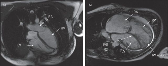
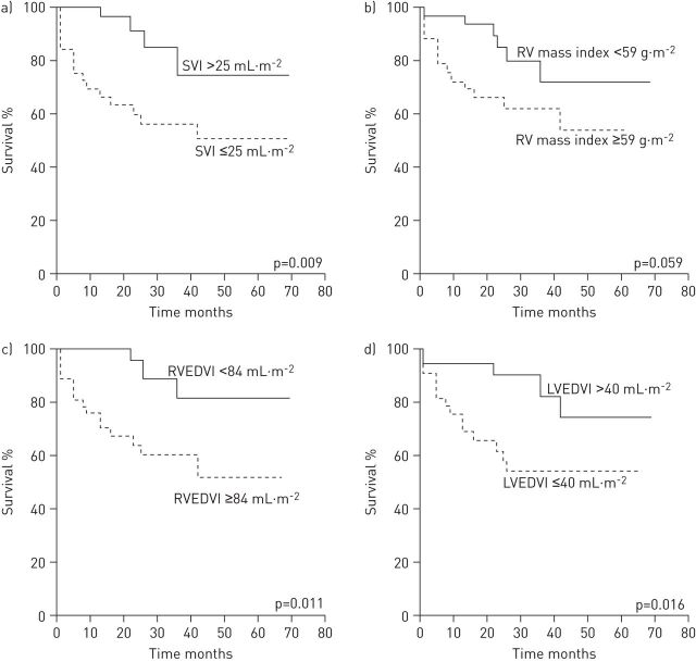
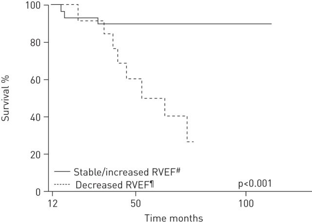
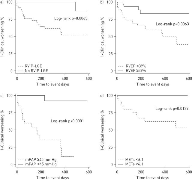
Cardiac magnetic resonance imaging (CMRI) provides accurate information about right ventricular (RV) mass, RV volumes and other markers of RV function. CMRI is proving to be a particularly useful tool in pulmonary arterial hypertension (PAH), as measures of RV function have been shown to be prognostic of long-term outcomes in this disease. Changes in RV function can also provide important information about a patient's disease course and response to treatment. As CMRI is noninvasive it can be used to regularly monitor patients with PAH, which is an important advantage over invasive right heart catheterisation. This review will explore the use of CMRI in the context of existing monitoring tools for PAH and will explore the forthcoming developments that are likely to be important in the future monitoring of patients with PAH.
Conflict of interest statement
Conflict of interest: Disclosures can be found alongside the online version of this article at
Figures
References
-
- Chin KM, Kim NH, Rubin LJ. The right ventricle in pulmonary hypertension. Coron Artery Dis 2005; 16: 13–18. - PubMed
-
- D'Alonzo GE, Barst RJ, Ayres SM, et al. Survival in patients with primary pulmonary hypertension. Results from a national prospective registry. Ann Intern Med 1991; 115: 343–349. - PubMed
-
- van Wolferen SA, van de Veerdonk MC, Mauritz GJ, et al. Clinically significant change in stroke volume in pulmonary hypertension. Chest 2011; 139: 1003–1009. - PubMed
-
- van Wolferen SA, Marcus JT, Boonstra A, et al. Prognostic value of right ventricular mass, volume, and function in idiopathic pulmonary arterial hypertension. Eur Heart J 2007; 28: 1250–1257. - PubMed
Publication types
MeSH terms
LinkOut - more resources
Full Text Sources
Other Literature Sources
Medical