. 2014 Mar;99(3):554-60.
doi: 10.3324/haematol.2013.093047. Epub 2013 Nov 29.

Loss of ATE1-mediated arginylation leads to impaired platelet myosin phosphorylation, clot retraction, and in vivo thrombosis formation

Affiliations

Loss of ATE1-mediated arginylation leads to impaired platelet myosin phosphorylation, clot retraction, and in vivo thrombosis formation

Lurong Lian et al. Haematologica. 2014 Mar.

Abstract

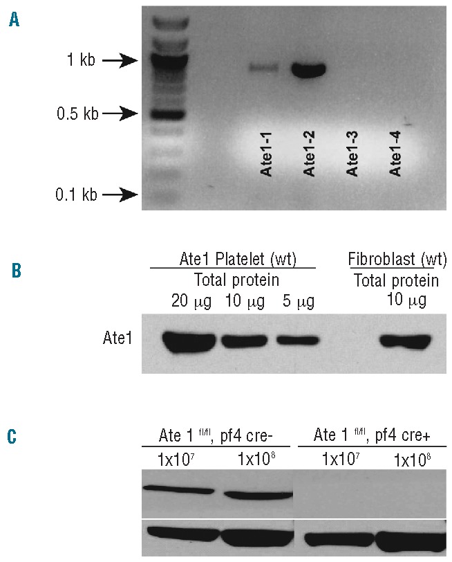
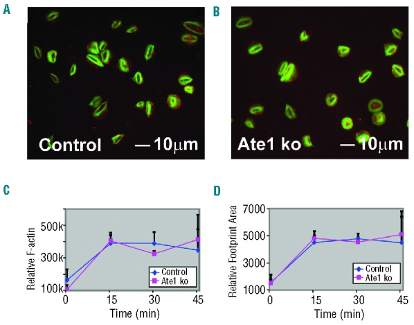
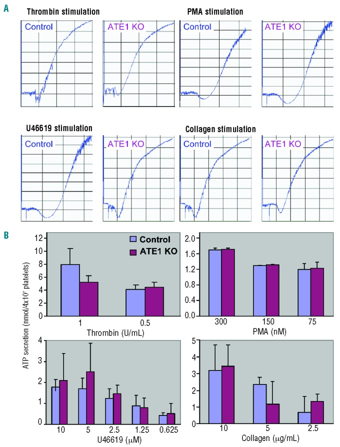
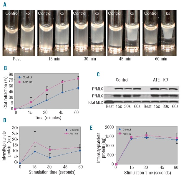
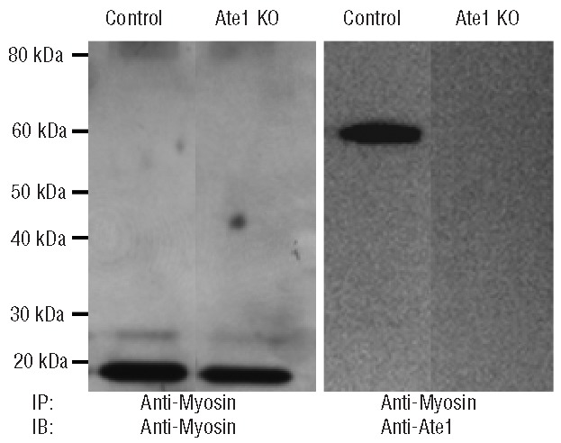
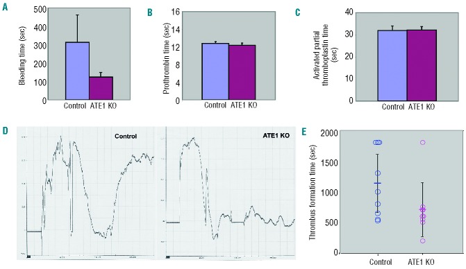
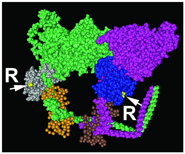
Protein arginylation by arginyl-transfer RNA protein transferase (ATE1) is emerging as a regulator protein function that is reminiscent of phosphorylation. For example, arginylation of β-actin has been found to regulate lamellipodial formation at the leading edge in fibroblasts. This finding suggests that similar functions of β-actin in other cell types may also require arginylation. Here, we have tested the hypothesis that ATE1 regulates the cytoskeletal dynamics essential for in vivo platelet adhesion and thrombus formation. To test this hypothesis, we generated conditional knockout mice specifically lacking ATE1 in their platelets and in their megakaryocytes and analyzed the role of arginylation during platelet activation. Surprisingly, rather than finding an impairment of the actin cytoskeleton structure and its rearrangement during platelet activation, we observed that the platelet-specific ATE1 knockout led to enhanced clot retraction and in vivo thrombus formation. This effect might be regulated by myosin II contractility since it was accompanied by enhanced phosphorylation of the myosin regulatory light chain on Ser19, which is an event that activates myosin in vivo. Furthermore, ATE1 and myosin co-immunoprecipitate from platelet lysates. This finding suggests that these proteins directly interact within platelets. These results provide the first evidence that arginylation is involved in phosphorylation-dependent protein regulation, and that arginylation affects myosin function in platelets during clot retraction.


Figures

Figure 1.
Figure 1.
ATE1 expression in mouse platelets and its abolishment in ATE1 knockout animals. (A) Reverse transcriptase polymerase chain reaction of RNA extracted from wild-type (WT) mouse platelets with ATE1 isoform-specific primer sets. ATE1-2 was the dominant isoform, while ATE1-3 and ATE1-4 were not detectable in platelets. (B) Comparisons of ATE1 levels in platelets and fibroblasts. (C) Immunoblot of the lysates from ATE1-deleted (Knockout) platelets or ATE1-floxed PF4 CRE (control) platelets verified the absence of ATE1 protein in the platelets of the mouse knockout model.
Figure 2.
Figure 2.
Platelet spreading on fibrinogen in response to thrombin. (A and B) Control and ATE1 knockout (ko) platelets were allowed to spread on fibrinogen-coated coverslips in the presence of thrombin. The cells were fixed and stained with phalloidin to detect F-actin (green) and antibodies were directed to detect β-actin (red). F-actin was concentrated centrally in the cortex of the spread platelets while β-actin was detected mostly at the edges. (C) Relative F-actin levels of platelets were assessed by flow cytometry staining with Alexa 488-Phalloidin. (D) The relative area (footprint size) of spread platelets is shown. The mean ± standard deviation are shown.
Figure 3.
Figure 3.
Platelet aggregation and secretion. (A) Aggregation tracings of platelets derived from control and ATE1 knockout (ko) mice after stimulation with various agonists. (B) Platelets derived from control or ATE1 KO platelets were analyzed for secretion of ATP using a luceriferase assay.
Figure 4.
Figure 4.
Increased clot retraction and phosphorylation of myosin in platelets lacking ATE1. (A) Platelet-rich plasma from ATE1 knockout (ko) and control mice was stimulated with thrombin at 37°C to induce clotting. Clot formation and retraction were continuously photographed before and after thrombin stimulation at 15 min intervals. Representative images at each time point show increased clot retraction in ATE1 ko platelets as compared to control platelets. (B) Quantification of clot retraction at each time point was measured as the percentage of the retracted area as compared to the initial total area of the clot (n=5.) Deletion of ATE1 significantly increased clot retraction (P<0.05, except at 60 min). The mean ± standard deviation are shown. (C) Phosphorylation of RLC at residues Ser19 and Thr18 was undetectable in resting platelets, but increased following stimulation with thrombin. Phosphorylation of RLC at either Ser19 (D) or Thr18 (E) was quantified based on the immunoblots of platelets stimulated with thrombin. The level of Ser19-RLC phosphorylation was significantly increased in platelets lacking ATE1 as compared to that in the controls (P<0.05) at 15 seconds (P<0.03) and at 30 seconds (P<0.05) after agonist stimulation. In contrast, Thr18-RLC phosphorylation remained the same in both groups. The mean ± standard deviation are shown.
Figure 5.
Figure 5.
ATE1 and myosin co-immunoprecipitate in murine platelets. Myosin was immunoprecipitated from lysates of ATE1 knockout (KO) and control platelets. The immunoprecipitates were fractionated by polyacrylamide gel electrophoresis and immunoblotted with myosin or ATE1 antibodies. On the left, an anti-myosin immunoblot demonstrates that myosin was equally immunoprecipitated from control and ATE1 knockout platelets. The immunoblot on the right demonstrates that the anti-myosin immunoprecipitates also contain ATE1.
Figure 6.
Figure 6.
Accelerated in vivo thrombus formation in mice with platelets that lack ATE1. (A, B, and C) Mice lacking ATE1 in their platelets had shorter tail bleeding times than their littermate controls (P<0.05), but the prothrombin time (PT) and activated partial thromboplastin time (aPTT) were identical in these two genotypes (P>0.05 for both PT and aPTT). (D) Carotid injury was induced by the application of FeCl3-soaked filter paper for 2 min, and arterial blood flow was monitored by a Doppler ultrasound probe. As shown by the representative tracings, ATE1 KO platelets were more likely to occlude faster and form stable thrombi. (E) The mean time of the thrombus formation is indicated by the short horizontal bar in the graph. Thrombus formation was quicker in the mice lacking ATE1 in their platelets (P=0.024).
Figure 7.
Figure 7.
RLC regulation of the 19th amino acid phosphorylation site by ATE1 arginylation of the essential light chain (ELC). Structural model of the myosin head and neck region (pink and green for two myosin heavy chain fragments) with the bound light chains (PDB identifier 1I84). The arginylated sites (denoted by yellow dots, arrows, and letters R) are found in the myosin ELC (blue and gray), adjacent to the RLC (brown and gold). It is likely that arginylation in these regions affects myosin conformation and/or binding of the enzymatic machinery that regulates myosin motor activity.

Comment in

References

    1. Kwon YT, Kashina AS, Davydov IV, Hu RG, An JY, Seo JW, et al. An essential role of N-terminal arginylation in cardiovascular development. Science. 2002;297(5578):96–9 - PubMed
    1. Rai R, Kashina A. Identification of mammalian arginyltransferases that modify a specific subset of protein substrates. Proc Natl Acad Sci USA. 2005;102(29):10123–8 - PMC - PubMed
    1. Carpio MA, Lopez Sambrooks C, Durand ES, Hallak ME. The arginylation-dependent association of calreticulin with stress granules is regulated by calcium. Biochem J. 2010;429(1):63. - PubMed
    1. Soffer RL. Enzymatic modification of proteins. V. Protein acceptor specificity in the arginine-transfer reaction. J Biol Chem. 1971;246(6):1602–6 - PubMed
    1. Ciechanover A, Ferber S, Ganoth D, Elias S, Hershko A, Arfin S. Purification and characterization of arginyl-tRNA-protein transferase from rabbit reticulocytes. Its involvement in post-translational modification and degradation of acidic NH2 termini substrates of the ubiquitin pathway. J Biol Chem. 1988;263(23):11155–67 - PubMed

Publication types