

Review
doi: 10.5665/sleep.3188.
The role of Hcrt/Orx and MCH neurons in sleep-wake state regulation
Affiliations
- PMID: 24293746
- PMCID: PMC3825421
- DOI: 10.5665/sleep.3188
Item in Clipboard
Review
The role of Hcrt/Orx and MCH neurons in sleep-wake state regulation
Sleep.
.
No abstract available
Figures
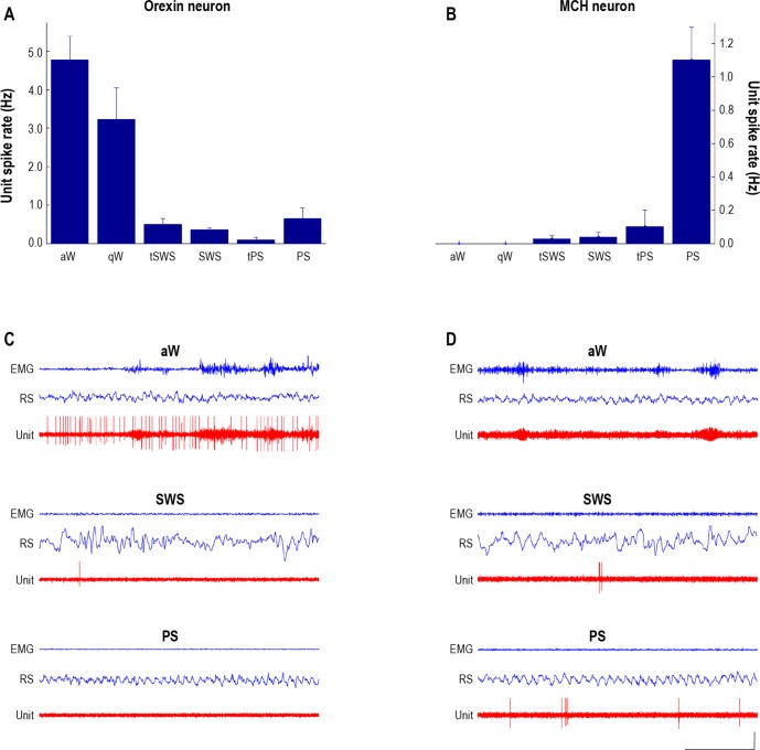
Discharge profiles of identified Orx and MCH neurons. Bar graphs showing the average spike rates (spikes per sec in Hz) of neurobiotin-labeled immunohistochemically identified Orx (A) and MCH (B) neurons along with (5 sec) periods of unit recordings with EEG and EMG (C and D, respectively). A and C: The Orx+ unit (C32u10) discharged maximally during active wake (aW) (average rate of 4.85 Hz). From aW, it decreased firing during quiet wake (qW), more significantly during transition to slow wave sleep (tSWS), to fire minimally during slow wave sleep (SWS), transition to paradoxical sleep (tPS) and paradoxical sleep (PS). Although the Orx unit was silent for long periods of PS (as illustrated), including periods of phasic twitching, it increased its firing with intervening periods of increased postural muscle tone and within several seconds prior to awakening (not shown). During maximal firing, it tended to fire in trains of spikes (with a mode interspike interval of 93.02 ms and corresponding instantaneous firing frequency of 10.75 Hz). B and D: The MCH+ unit (C134u01) discharged maximally during PS (1.1 Hz). It was silent during both aW and qW, fired at very low rates during tSWS, SWS, and tPS and at its highest (albeit still low) rate during PS. During maximal firing, it tended to fire very phasically with doublets or clusters of spikes (a mode interspike interval of 17.01 ms and corresponding instantaneous firing frequency of 58.75 Hz). The discharge rates of the Orx and MCH units were respectively (and reciprocally) positively and negatively correlated with EMG amplitude across the sleep-waking cycle. Calibrations in C and D: horizontal, 1 sec; vertical, 1 mV (EEG, EMG), 2 mV (unit). Adapted with permission from Lee and Hassani.
Comment in
-
Relation of melanin concentrating hormone levels to sleep, emotion and hypocretin levels.Sleep. 2013 Dec 1;36(12):1777. doi: 10.5665/sleep.3194. Sleep. 2013. PMID: 24293749 Free PMC article. No abstract available.
References
-
- Modirrousta M, Mainville L, Jones BE. Orexin and MCH neurons express c-Fos differently after sleep deprivation vs. recovery and bear different adrenergic receptors. Eur J Neurosci. 2005;21:2807–16. - PubMed
Publication types
MeSH terms
Substances
LinkOut - more resources
Full Text Sources
Other Literature Sources
