. 2013 Dec 1;36(12):1849-58.
doi: 10.5665/sleep.3214.

Intermittent hypoxia exacerbates pancreatic β-cell dysfunction in a mouse model of diabetes mellitus

Affiliations

Intermittent hypoxia exacerbates pancreatic β-cell dysfunction in a mouse model of diabetes mellitus

Shariq I Sherwani et al. Sleep. .

Abstract

Study objectives: The effects of intermittent hypoxia (IH) on pancreatic function in the presence of diabetes and the underlying mechanisms are unclear. We hypothesized that IH would exacerbate pancreatic β-cell dysfunction and alter the fatty acids in the male Tallyho/JngJ (TH) mouse, a rodent model of type 2 diabetes.

Design: TH mice were exposed for 14 d to either 8 h of IH or intermittent air (IA), followed by an intraperitoneal glucose tolerance test (IPGTT) and tissue harvest. The effect of IH on insulin release was determined by using a β3-adrenergic receptor (AR) agonist.

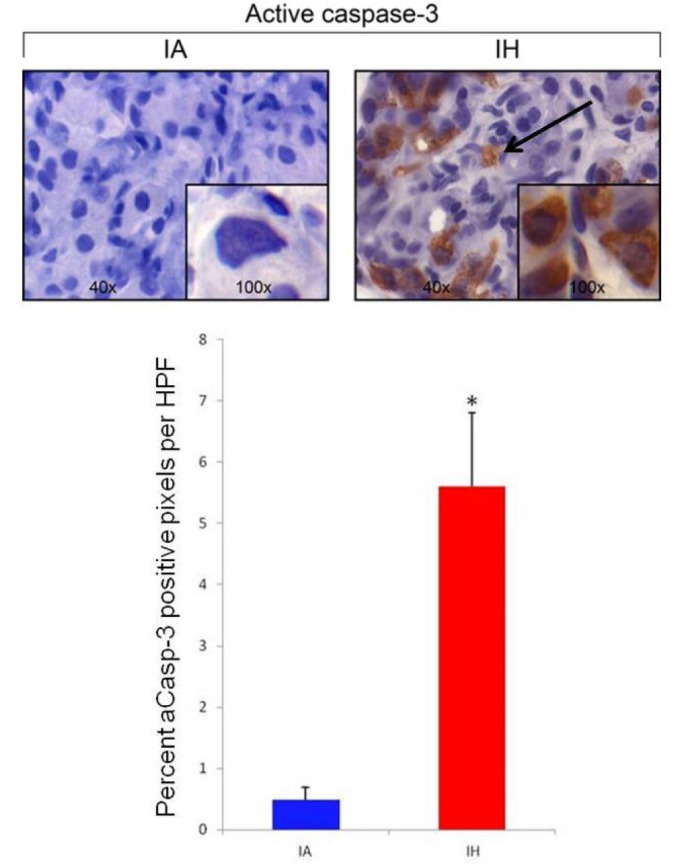
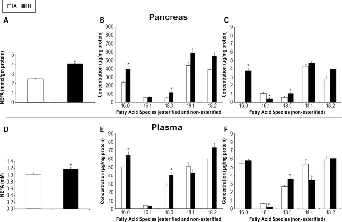
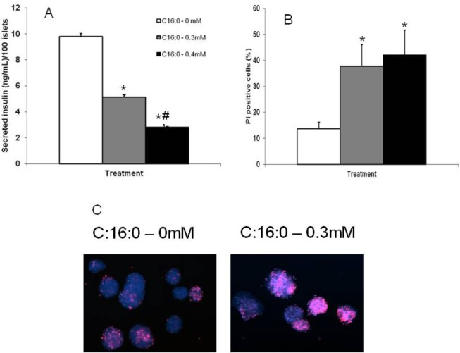
Measurements and results: During IH, pancreatic tissue pO2 decreased from 20.4 ± 0.9 to 5.7 ± 2.6 mm Hg, as determined by electron paramagnetic resonance oximetry. TH mice exposed to IH exhibited higher plasma glucose levels during the IPGTT (P < 0.001) while the insulin levels tended to be lower (P = 0.06). Pancreatic islets of the IH group showed an enhancement of the caspase-3 staining (P = 0.002). IH impaired the β-AR agonist-mediated insulin release (P < 0.001). IH increased the levels of the total free fatty acids and saturated fatty acids (palmitic and stearic acids), and decreased levels of the monounsaturated fatty acids in the pancreas and plasma. Ex vivo exposure of pancreatic islets to palmitic acid suppressed insulin secretion and decreased islet cell viability.

Conclusions: Intermittent hypoxia increases pancreatic apoptosis and exacerbates dysfunction in a polygenic rodent model of diabetes. An increase in free fatty acids and a shift in composition towards long chain saturated fatty acid species appear to mediate these effects.

Keywords: Intermittent hypoxia; diabetes mellitus; fatty acids; β-cell function.


Figures

Figure 1
Figure 1
Changes in the FiO2 (A) during a cycle of the IH stimulus used in our experiments and the corresponding pancreatic tissue pO2 (B) measured by electron paramagnetic resonance (EPR) oximetry (n = 3). At baseline the pancreatic tissue pO2 was 20.4 ± 0.9 mm Hg. During the IH cycle pancreatic tissue pO2 nadir was 5.7 ± 2.6 mm Hg.
Figure 2
Figure 2
IPGTT in TH mice. TH mice exposed to IH (n = 6) had significantly higher plasma glucose values compared to the TH mice exposed to IA (n = 6) and the multiple comparison procedure showed that the values were different at all time periods. The IH exposed mice had lower insulin levels compared to the IA exposed mice but the analysis did not achieve statistical significance when all time points were included (P = 0.06). The AUC analysis is shown in the right panel. The AUC of the glucose levels was significantly higher in the IH exposed mice (P < 0.001). Despite the higher glucose values, the AUC of the insulin levels was lower in the TH mice exposed to IH, although this did not reach statistical significance (P = 0.056). The results obtained in the experiments (n = 12) wherein the IPGTT was performed under anesthesia were similar except that the insulin values were significantly lower in the TH mice exposed to IH (see supplemental material). *P < 0.05
Figure 3
Figure 3
Pancreatic islet active caspase 3 (aCasp-3) IHC. After the IPGTT, TH mice were sacrificed and their pancreas harvested. TH mice exposed to IH (n = 6) had increased aCasp-3 brown staining (arrow) compared to IA exposed mice (n = 6) indicating increased apoptosis in pancreatic islet cells *P = 0.002.
Figure 4
Figure 4
β3-AR agonist sensitivity test. In the non-fasted state, TH mice exposed to IH (n = 6) showed an impaired β-AR agonist-mediated insulin release compared to the TH mice exposed to IA (n = 6). The plasma insulin levels of the IH group were lower compared to the IA group at all time points after the IP injection of 1 mg/kg CL 316,243, a selective β3-AR agonist. Overall, TH mice exposed to IH also exhibited an impaired β3-AR agonist-induced release of NEFAs. A multiple comparison procedure showed that the NEFA levels were significantly different between groups only at 60 min after the IP injection of CL 316,243. *P < 0.05
Figure 5
Figure 5
Effects of IH on fatty acids. (A) IH exposure caused a significant increase in the total NEFA levels in the pancreas (P = 0.002). (B) Analysis of the fatty acid species of the total (esterified and non-esterified) fatty acids showed that the long chain saturated fatty acids, palmitic (C16:0) and stearic (C18:0), were significantly increased in IH exposed mice (P < 0.001 and 0.009, respectively). (C) Similar results were obtained in the analysis of the fatty acid species of the NEFAs in the pancreas (P = 0.007 and 0.009, respectively). Additionally, in the NEFA fraction, IH also caused a significant decrease in the monounsaturated fatty acid, palmitoleic (C16:1) (P = 0.009). (D) IH exposure caused a significant increase in the total NEFA levels in the plasma (P = 0.04). (E) Analysis of the fatty acid species of the total (esterified and non-esterified) fatty acids showed that the long chain saturated fatty acids, palmitic (C16:0) and stearic (C18:0), were significantly increased in IH exposed mice (P = 0.011 and 0.005, respectively). (F) Analysis of the fatty acid species of the NEFAs in the plasma showed that IH caused a significant increase only in stearic (C18:0) acid (P = 0.004), while the tendency for an increased circulating palmitic (C16:0) acid did not achieve statistical significance. Additionally, in the NEFA fraction, IH also caused a significant decrease in the monounsaturated fatty acids, palmitoleic (C16:1) and oleic (C18:1) (P < 0.001 and 0.007, respectively).
Figure 6
Figure 6
Ex vivo studies. Pancreatic islets were isolated from TH mice and the isolated islets were exposed to different concentrations of the long chain unsaturated FA, palmitic acid (C16:0). (A) After 24 h of incubation with C16:0, secreted insulin level was significantly reduced (P < 0.001). (B) Exposure to C16:0 caused a significant increase in islet cell death (P = 0.002). (C) Representative images are shown that illustrate the increased cell death seen in islets incubated with C16:0 compared to control. *Significantly different from control, P < 0.05; #significantly different from 0.3 mM condition, P < 0.05.

References

    1. Ip MS, Lam B, Ng MM, Lam WK, Tsang KW, Lam KS. Obstructive sleep apnea is independently associated with insulin resistance. Am J Respir Crit Care Med. 2002;165:670–6. - PubMed
    1. Punjabi NM, Shahar E, Redline S, Gottlieb DJ, Givelber R, Resnick HE. Sleep-disordered breathing, glucose intolerance, and insulin resistance: the Sleep Heart Health Study. Am J Epidemiol. 2004;160:521–30. - PubMed
    1. Punjabi NM, Beamer BA. Alterations in glucose disposal in sleep-disordered breathing. Am J Respir Crit Care Med. 2009;179:235–40. - PMC - PubMed
    1. Seicean S, Kirchner HL, Gottlieb DJ, et al. Sleep-disordered breathing and impaired glucose metabolism in normal-weight and overweight/obese individuals: the Sleep Heart Health Study. Diabetes Care. 2008;31:1001–6. - PubMed
    1. Brooks B, Cistulli PA, Borkman M, et al. Obstructive sleep apnea in obese noninsulin-dependent diabetic patients: effect of continuous positive airway pressure treatment on insulin responsiveness. J Clin Endocrinol Metab. 1994;79:1681–5. - PubMed

Publication types