. 2013:8:4481-94.
doi: 10.2147/IJN.S41782. Epub 2013 Nov 21.

Targeted delivery of let-7a microRNA encapsulated ephrin-A1 conjugated liposomal nanoparticles inhibit tumor growth in lung cancer

Affiliations

Targeted delivery of let-7a microRNA encapsulated ephrin-A1 conjugated liposomal nanoparticles inhibit tumor growth in lung cancer

Hung-Yen Lee et al. Int J Nanomedicine. 2013.

Abstract

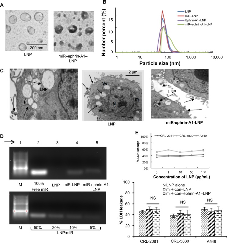
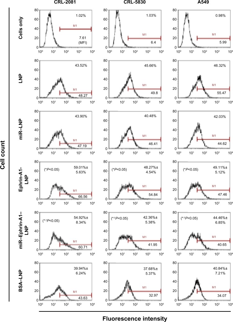
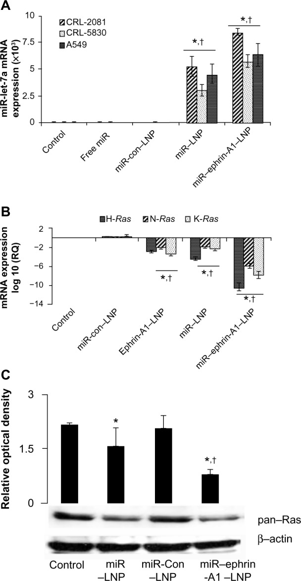
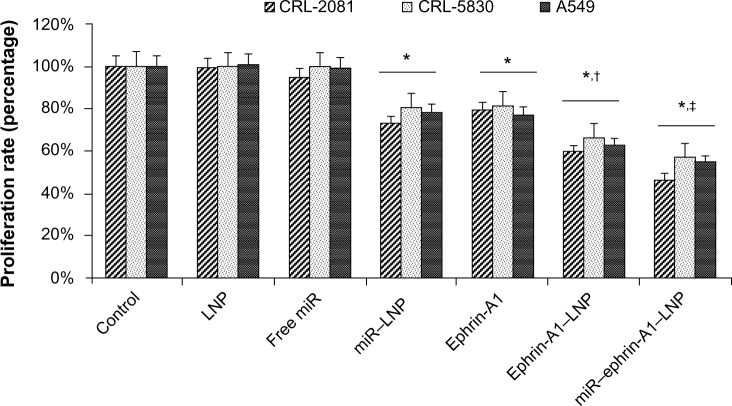
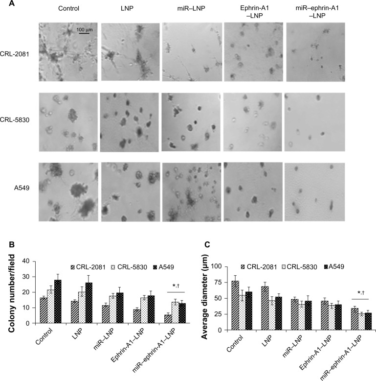
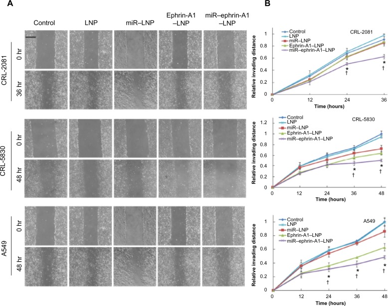
MicroRNAs (miRs) are small noncoding RNA sequences that negatively regulate the expression of target genes by posttranscriptional repression. miRs are dysregulated in various diseases, including cancer. let-7a miR, an antioncogenic miR, is downregulated in lung cancers. Our earlier studies demonstrated that let-7a miR inhibits tumor growth in malignant pleural mesothelioma (MPM) and could be a potential therapeutic against lung cancer. EphA2 (ephrin type-A receptor 2) tyrosine kinase is overexpressed in most cancer cells, including MPM and non-small-cell lung cancer (NSCLC) cells. Ephrin-A1, a specific ligand of the EphA2 receptor, inhibits cell proliferation and migration. In this study, to enhance the delivery of miR, the miRs were encapsulated in the DOTAP (N-[1-(2.3-dioleoyloxy)propyl]-N,N,N-trimethyl ammonium)/Cholesterol/DSPE (1,2-distearoyl-sn-glycero-3-phosphoethanolamine-N-[cyanur(polyethylene glycol)-2000])-PEG (polyethylene glycol)-cyanur liposomal nanoparticles (LNP) and ephrin-A1 was conjugated on the surface of LNP to target receptor EphA2 on lung cancer cells. The LNP with an average diameter of 100 nm showed high stability, low cytotoxicity, and high loading efficiency of precursor let-7a miR and ephrin-A1. The ephrin-A1 conjugated LNP (ephrin-A1-LNP) and let-7a miR encapsulated LNP (miR-LNP) showed improved transfection efficiency against MPM and NSCLC. The effectiveness of targeted delivery of let-7a miR encapsulated ephrin-A1 conjugated LNP (miR-ephrin-A1-LNP) was determined on MPM and NSCLC tumor growth in vitro. miR-ephrin-A1-LNP significantly increased the delivery of let-7a miR in lung cancer cells when compared with free let-7a miR. In addition, the expression of target gene Ras was significantly repressed following miR-ephrin-A1-LNP treatment. Furthermore, the miR-ephrin-A1-LNP complex significantly inhibited MPM and NSCLC proliferation, migration, and tumor growth. Our results demonstrate that the engineered miR-ephrin-A1-LNP complex is an effective carrier for the targeted delivery of small RNA molecules to lung cancer cells. This could be a potential therapeutic approach against tumors overexpressing the EphA2 receptor.

Keywords: EphA2 receptor; ephrin-A1; liposomal nanoparticles; malignant pleural mesothelioma; microRNA; non-small-cell lung cancer.


Figures

Figure 1
Figure 1
Characterization of engineered LNPs. Notes: (A) Transmission electron microscopy image of LNPs. (B) Particle size distribution of LNP and miR–encapsulated LNP, ephrin-A1 conjugated LNP, and miR–ephrin-A1–LNP. (C) Cellular uptake of LNP as determined by transmission electron microscopy. The left hand panel is at a higher magnification. Arrows point to LNP with vesicles; bar represents 2 μm, middle panel. (D) The encapsulation of miR in LNPs was determined by using gel electrophoresis. In order to compare the encapsulation of miR, free miR (100% miR, upper panel) is shown versus 50%, 20%, 10%, and 5% of miR that encapsulated with LNPs, lower panel. The encapsulation efficiency of miR in LNPs was around 90% and remained high after protein conjugation; arrow points to the lane number in the gel. (E) LDH leakage after incubation of lung cancer cells with LNPs. Engineered LNPs showed low cytotoxicity on all three lung cancer cells tested, upper panel. LNP at an even higher concentration (100 μg/mL) failed to show any significant cytotoxicity in CRL-2081, CRL-5830, and A549 cells. The encapsulation of miR–ephrin-A1–LNP and ephrin-A1–LNP did not induce any cytotoxic effect on the cells, lower panel. Data represents mean ± SEM of three separate experiments. Abbreviations: Con, control; LDH, lactate dehydrogenase; LNP, liposomal nanoparticle; m, mitochondria; M, marker; miR, microRNA; N, nucleus; NS, not significant; SEM, standard error of the mean; Vs, vesicles.
Figure 2
Figure 2
Cellular uptake of LNP and protein conjugated LNP in lung cancer cells as determined by flow cytometry analysis. Notes: Cellular uptake of LNPs was evaluated after 40 minutes of incubation of cell cultures with LNP. The cellular uptake efficiency of the cationic LNPs was high in all the lung cancer cells (CRL-2081, CRL-5830 and A549 cells). The ephrin-A1 conjugation on the LNP surface showed enhanced cellular uptake via ligand targeting on lung cancer cells. The ephrin-A1–LNP showed the highest percent cellular uptake and intensity in CRL-2081 cells when compared with CRL-5830 and A549 cell lines. However, ephrin-A1 conjugation on the LNPs enhanced the uptake efficiency compared with the LNPs conjugated with nonspecific proteins (BSA–LNP). miR–ephrin-A1–LNP cellular was significantly higher when compared with miR–LNP. Data presented is mean ± SEM of three independent experiments. *P<0.05 when compared with BSA–LNP; P<0.05 when compared with miR–LNP alone. Abbreviations: BSA, bovine serum albumin; LNP, liposomal nanoparticle; miR, microRNA; SEM, standard error of the mean.
Figure 3
Figure 3
miR–LNP and miR–ephrin-A1–LNP complexes enhance let-7a miR expression in lung cancer cells. Notes: (A) CRL-2081, CRL-5830, and A549 cells were transfected with miR–ephrin-A1–LNP, miR–LNP, miR-con–LNP, or free miR, or were left untransfected (control) for 24 hours. The let-7a miR mRNA expression was measured by quantitative real-time PCR. Data is presented as the relative quantification compared with control (cells cultured in medium alone) samples as the baseline value. Data represent the mean ± SEM of three separate experiments. 18S RNA was probed as endogenous control to normalize the expression of let-7a miR. *P<0.001, cells transfected with miR–ephrin-A1–LNP versus control; P<0.001 when compared with miR-con–LNP. (B) H-Ras, N-Ras, and K-Ras mRNA expression as analyzed by quantitative real-time PCR. CRL-2081 were transfected with miR–ephrin-A1–LNP, miR–LNP, ephrin-A1–LNP, or miR-con–LNP, or were left untransfected (control) for 24 hours. Data presented are the mean ± SEM of three separate experiments. 18S RNA was used as endogenous control to normalize the expression of let-7a miR. *P<0.001, CLR-2081 transfected with miR–ephrin-A1–LNP versus control; P<0.05 when compared with miR-con–LNP. (C) Ras expression in CRL-2081 as detected by Western blot analysis. CRL-2081 were transfected with miR–ephrin-A1–LNP, miR-con–LNP, miR–LNP, or left untransfected to determine ras expression. The β-actin was detected to demonstrate equal loading of protein. Upper panel represents relative optical density. Data presented are the mean ± SEM of three separate experiments. *P<0.001, CLR-2081 transfected with miR–ephrin-A1–LNP versus control; P<0.05 when compared with miR-con–LNP. Abbreviations: con, control; LNP, liposomal nanoparticle; miR, microRNA; PCR, polymerase chain reaction; RQ, relative quantification; SEM, standard error of the mean.
Figure 4
Figure 4
Effect of LNP complexes on lung cancer cell proliferation. Notes: Lung cancer cell lines CRL-2081, CRL-5830, and A549 were treated with LNP complexed with miR or ephrin-A1 and compared with free miR and ephrin-A1 after 48 hours of treatment. The miR–ephrin-A1–LNP showed enhanced inhibitory effect on cell proliferation. Data presented is mean ± SEM of three separate experiments. *P<0.05 when compared with control; P<0.05 when compared with LNP; P<0.05 when compared with miR–LNP complex. Abbreviations: Con, control; LNP, liposomal nanoparticle; miR, microRNA; PCR, polymerase chain reaction; RQ, relative quantification; SEM, standard error of the mean.
Figure 5
Figure 5
The LNP complexes reduce lung cancer tumor growth in three-dimensional Matrigels in vitro. Notes: The lung cancer cells were incubated in serum free medium (control) or treated either with ephrin-A1–LNP or miR–ephrin-A1–LNP or LNP alone, and tumor formation was determined in Matrigel. (A) The miR–ephrin-A1–LNP was highly effective on tumor growth inhibition of CRL-2081, CRL-5830 and A549 cells in Matrigel. (B and C) The quantification data of tumor colonies and size. The data presented are the mean ± SEM of three independent experiments performed at separate times; values were considered significant at *P<0.001 when compared with control and P<0.001 when compared with miR–LNP complex and miR–ephrin-A1–LNP. Abbreviations: LNP, liposomal nanoparticle; miR, microRNA.
Figure 6
Figure 6
LNP complexes inhibit invasion of lung cancer cells. Notes: Lung cancer cell invasion was determined by wound healing assay. (A) Optical images of wounded cell monolayers were taken at 0 hours and 36 hours (CRL-2081) or 48 hours (CRL-5830 and A549) after wounding the cultures. The cell migration rates of CRL-2081, CRL-5830, and A549 were determined by measuring the distance cells invaded over time from the edge of wound. (B) The invading distances are shown in relative distance to the control cultures (relative invading distance = invading distance of sample/invading distance of control sample; relative invading distance of control sample is 1). The miR–ephrin-A1–LNP complex effectively reduced the recovery rate of wound area on the monolayer. The data presented is the mean ± SEM of three independent experiments performed at separate times; values were considered significant at *P<0.001 when compared with control and P<0.001 when compared with miR–LNP complex and miR–ephrin-A1–LNP. Abbreviations: LNP, liposomal nanoparticle; miR, microRNA; SEM, standard error of the mean.

References

    1. Lee RC, Feinbaum RL, Ambros V. The C elegans heterochronic gene lin-4 encodes small RNAs with antisense complementarity to lin-14. Cell. 1993;75(5):843–854. - PubMed
    1. Bartel DP. MicroRNAs: genomics, biogenesis, mechanism, and function. Cell. 2004;116(2):281–297. - PubMed
    1. Reinhart BJ, Slack FJ, Basson M, et al. The 21-nucleotide let-7 RNA regulates developmental timing in Caenorhabditis elegans. Nature. 2000;403(6772):901–906. - PubMed
    1. Khodayari N, Mohammed KA, Goldberg EP, Nasreen N. EphrinA1 inhibits malignant mesothelioma tumor growth via let-7 microRNA-mediated repression of the RAS oncogene. Cancer Gene Ther. 2011;18(11):806–816. - PubMed
    1. Miao H, Wei BR, Peehl DM, et al. Activation of EphA receptor tyrosine kinase inhibits the Ras/MAPK pathway. Nat Cell Biol. 2001;3(5):527–530. - PubMed

Publication types