Randomized Controlled Trial
. 2013:8:4507-20.
doi: 10.2147/IJN.S53032. Epub 2013 Nov 21.

Cerium oxide nanoparticles protect rodent lungs from hypobaric hypoxia-induced oxidative stress and inflammation

Affiliations
Randomized Controlled Trial

Cerium oxide nanoparticles protect rodent lungs from hypobaric hypoxia-induced oxidative stress and inflammation

Aditya Arya et al. Int J Nanomedicine. 2013.

Abstract

Background: Cerium oxide nanoparticles (nanoceria) are effective at quenching reactive oxygen species (ROS) in cell culture and animal models. Although nanoceria reportedly deposit in lungs, their efficacy in conferring lung protection during oxidative stress remains unexplored. Thus, the study evaluated the protective efficacy of nanoceria in rat lung tissue during hypobaric hypoxia.

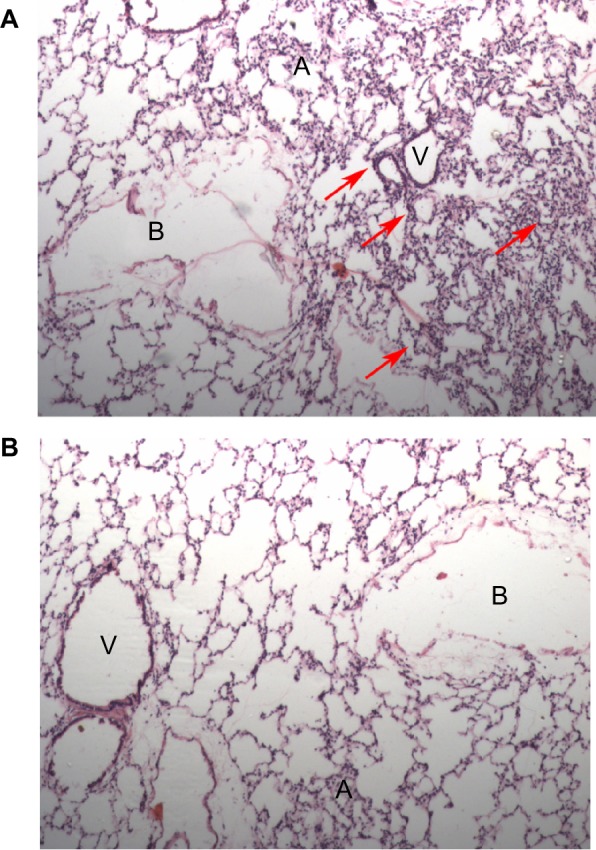
Methods: A total of 48 animals were randomly divided into four equal groups (control [C], nanoceria treated [T], hypoxia [H], and nanoceria treated plus hypoxia [T+H]). Animals were injected intraperitoneally with either a dose of 0.5 μg/kg body weight/week of nanoceria (T and T+H groups) or vehicle (C and H groups) for 5 weeks. After the final dose, H and T+H animals were challenged with hypobaric hypoxia, while C and T animals were maintained at normoxia. Lungs were isolated and homogenate was obtained for analysis of ROS, lipid peroxidation, glutathione, protein carbonylation, and 4-hydroxynonenal-adduct formation. Plasma was used for estimating major inflammatory cytokines using enzyme-linked immunosorbent assay. Intact lung tissues were fixed and both transmission electron microscopy and histopathological examinations were carried out separately for detecting internalization of nanoparticles as well as altered lung morphology.

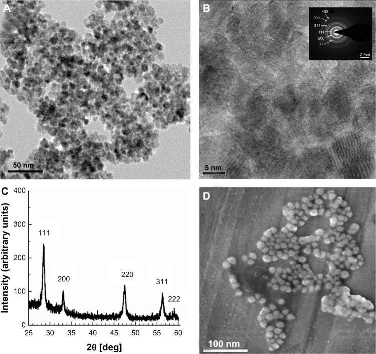
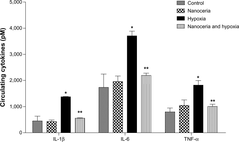
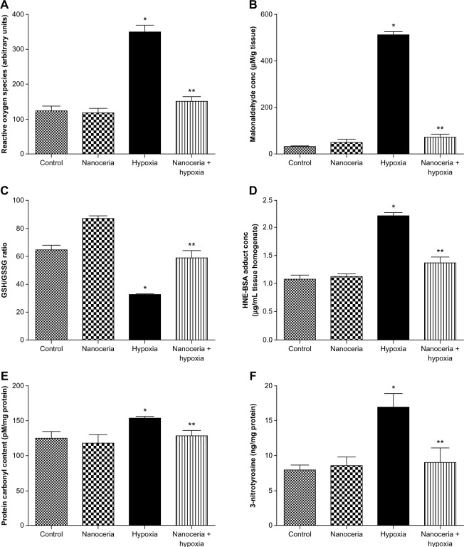
Results: Spherical nanoceria of 7-10 nm diameter were synthesized using a microemulsion method, and the lung protective efficacy of the nanoceria evaluated during hypobaric hypoxia. With repeated intraperitoneal injections of low micromole concentration, we successfully localized the nanoceria in rodent lung without any inflammatory response. The lung-deposited nanoceria limited ROS formation, lipid peroxidation, and glutathione oxidation, and prevented oxidative protein modifications like nitration and carbonyl formation during hypobaric hypoxia. We also observed reduced lung inflammation in the nanoceria-injected lungs, supporting the anti-inflammatory properties of nanoceria.

Conclusion: Cumulatively, these results suggest nanoceria deposit in lungs, confer protection by quenching noxious free radicals during hypobaric hypoxia, and do not evoke any inflammatory response.

Keywords: high altitude; nanoceria; nanomedicine.


Figures

Figure 1
Figure 1
Characterization of nanoceria. (A) High-resolution transmission electron microscopy (HR-TEM) photomicrograph revealing the spherical shape of nanoceria; the average diameter of each particle was 7–10 nm. (B) HR-TEM micrograph of nanoceria showing the crystalline fringes in particles. (Inset) The selected-area electron diffraction pattern further confirms the crystalline structure of the nanoceria. (C) X-ray diffraction of nanoceria particles showing the characteristic peaks of the crystal. The Miller indices demonstrate the high purity of the synthesized nanoparticles. (D) Scanning electron microscopy analysis showing the spherical shape and homogenous surface topology of the nanoparticles.
Figure 2
Figure 2
Lung localization of nanoceria. Deposition of electron-dense particles with a size of 7–10 nm in the alveolar spaces. The selected-area electron diffraction pattern (inset) further confirms the presence of nanoceria. Abbreviations: PM, plasma membrane; C, cytoplasm; N, nucleus.
Figure 3
Figure 3
Estimation of circulating cytokines in plasma. Concentrations of interleukin (IL)-1β (control [C]: 454.50±176.00, nanoceria treated [T]: 425.70±62.60, hypoxia [H]: 1,366.70±9.01, nanoceria treated plus hypoxia [T+H]: 548.88±33.60), IL-6 (C: 1,736.00±509.00, T: 1,962.60±212.22, h: 3,709.50±176.15, T+H: 2,189.00±83.87), and tumor necrosis factor (TNF)-α (C: 797.39±149.70, T:1,047.15±12.20, H: 1,813.20±76.10, T+H:1,005.70±83.80) were elevated during hypoxia, whereas animals administered nanoceria showed no such increases. The concentration of circulating cytokines in nanoceria-treated and hypoxia-exposed animals (n= 10) was significantly lower than that in hypoxic animals. Notes: *P<0.05; **P<0.01.
Figure 4
Figure 4
Histopathological examination of rat lung tissue for tissue inflammation. Photomicrographs of hematoxylin and eosin-stained lung sections (20×): (A) hypoxic lung sections showed localized neutrophil infiltration and inflammation (indicated by arrows); (B) nanoceria-deposited lungs did not show any sign of inflammation. Abbreviations: A, alveoli; B, bronchioles; V, blood vessels.
Figure 5
Figure 5
Estimation of oxidative stress indicators in lung tissue homogenate. (A) Hypoxic lung tissue showed an increase in the radical generation, while nanoceria-deposited lung exposed to hypoxia did not show any increase, as compared with the control. Nanoceria alone did not cause any increase in free radicals (control [C]: 123.8±13.4, nanoceria treated [T]: 119.3±11.2, hypoxia [H]: 350.2±19.2, nanoceria treated plus hypoxia [T+H]: 150.2±13.2). (B) Hypoxic lung showed an increase in lipid peroxidation, while nanoceria-deposited lung exposed to hypoxia did not show any increase in malonaldehyde levels (C: 30.9±5.3, T: 51.2±11.9, H: 512.3±13.3, T+H: 72.2±11.9). (C) The glutathione (GSH)/glutathione disulfide (GSSG) ratio was reduced in hypoxic lungs, while nanoceria-deposited lungs exposed to hypoxia had a GSH/GSSG ratio equivalent to that of the control. The GSH/GSSG ratio was increased in nanoceria alone (C: 65.0±2.8, T: 70.3±11.6, H: 32.4±0.4, T+H: 58.9±4.8). (D) In hypoxic lungs, 4-hydroxynoneal was increased, while it was stabilized in nanoceria-deposited lungs (C: 1.00±0.07, T: 1.10±0.05, H: 2.20±0.05, T+H: 1.30±0.09). (E) Protein carbonyl content was elevated in hypoxic lungs and restored in nanoceria-deposited lungs (C: 125.3±9.5, T: 118.6±11.3, H: 153.4±2.5, T+H: 128.6±8.2). (F) The concentration of 3-nitrotyrosine was increased during hypoxia, while no significant elevation was observed in nanoceria-deposited lungs exposed to hypoxia (C: 1.07±0.07, T: 1.10±0.05, H: 2.20±0.05, T+H: 1.30±0.09). Notes: All results are the means of three sets of experiments; error bars represent the standard error of the mean (n=10). *P<0.05; **P<0.01. Abbreviations: HNE, 4-hydroxynoneal; BSA, bovine serum albumin.

References

    1. Karakoti AS, Monteiro-Riviere NA, Aggarwal R, et al. Nanoceria as antioxidant: synthesis and biomedical applications. JOM (1989) 2008;60(3):33–37. - PMC - PubMed
    1. Heckert EG, Karakoti AS, Seal S, Self WT. The role of cerium redox state in the SOD mimetic activity of nanoceria. Biomaterials. 2008;29(18):2705–2709. - PMC - PubMed
    1. Pirmohamed T, Dowding JM, Singh S, et al. Nanoceria exhibit redox state-dependent catalase mimetic activity. Chem Commun (Camb) 2010;46(16):2736–2738. - PMC - PubMed
    1. Hirst SM, Karakoti A, Singh S, et al. Bio-distribution and in vivo antioxidant effects of cerium oxide nanoparticles in mice. Environ Toxicol. 2013;28(2):107–118. - PubMed
    1. Yokel RA, Au TC, MacPhail R, et al. Distribution, elimination, and biopersistence to 90 days of a systemically introduced 30 nm ceria-engineered nanomaterial in rats. Toxicol Sci. 2012;127(1):256–268. - PubMed

Publication types