

Short-term vasculoprotective effects of imatinib mesylate on intimal hyperplasia of arterial anastomosis: An experimental study using a rabbit model
- PMID: 24294014
- PMCID: PMC3513249
- DOI: 10.1177/229255031202000414
Short-term vasculoprotective effects of imatinib mesylate on intimal hyperplasia of arterial anastomosis: An experimental study using a rabbit model
Abstract
Background: Since the beginning of the 'microvascular era', the success rates of microvascular procedures have increased to more than 90% in most series. The main reason for failure, however, is the healing of microarterial anastomosis, which is dependent on the status of endothelial cells and affects the rate of arterial thrombosis. In 80% of arterial thrombosis cases, complications are primarily observed during the first 72 h after surgery. Healing of arterial anastomosis results in intimal hyperplasia in which myofibroblasts comprise the predominant cell type. Intimal hyperplasia has been described previously as an adaptive process that occurs in response to hemodynamic stress or injuries to the vascular bed. During wound healing, fibroblasts proliferate, migrate and differentiate into myofibroblasts - a process that takes one to three days. Imatinib mesylate (ST1571-Gleevec, Novartis, Germany) is a specific platelet-derived growth factor receptor blocker that has found use as an adjunct to sirolimus in cardiovascular surgery for restenosis. However, its potential utility in preventing arterial thrombosis in microvascular surgery has not been evaluated in routine plastic surgery practice.
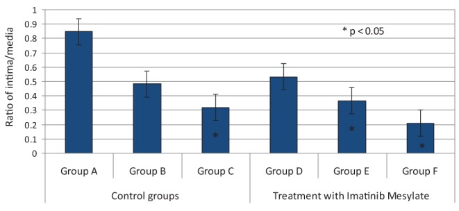
Methods: Twenty-four randomly selected, male, white New Zealand rabbits were divided into six groups (A to F), and the femoral artery model was used for arterial anastomosis. Following anastomosis, groups A, B and C received phosphate-buffered saline orogastrically. In groups D, E and F, imatinib mesylate was administered via an orogastric tube twice per day at a dose of 10 mg/kg starting two days before arterial anastomosis. Following anastomosis, imatinib mesylate was administered for one, three and seven days, and the regression of intimal hyperplasia was recorded.
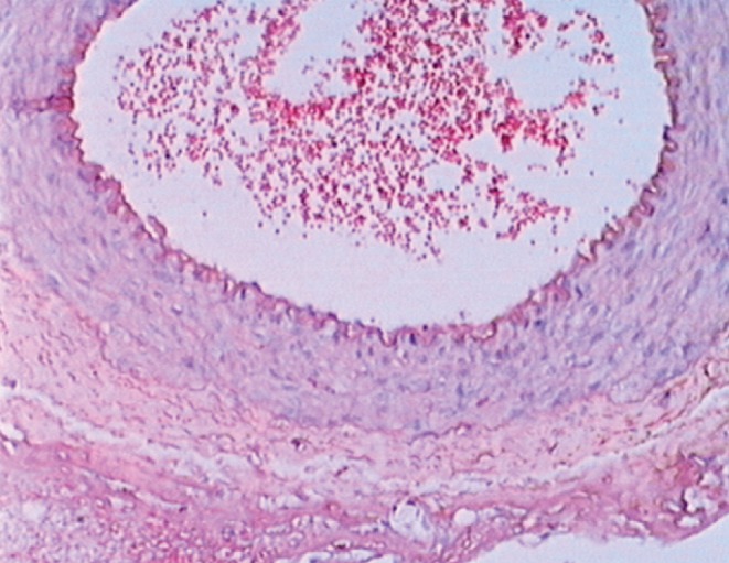
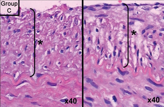
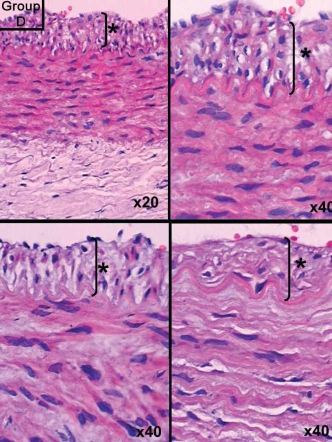
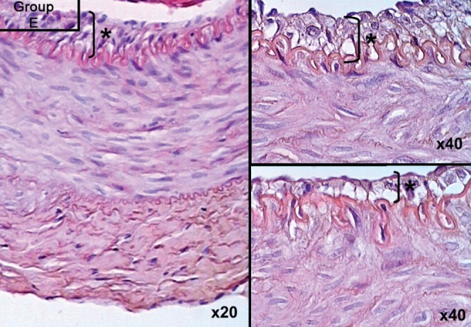
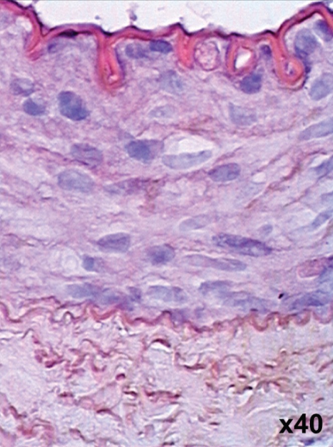
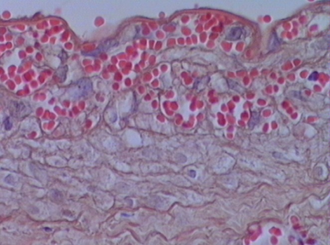
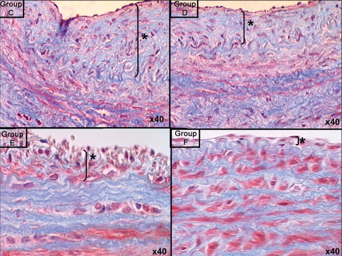
Results: In groups administered imatinib mesylate (ie, groups D, E and F), intimal hyperplasia decreased by up to 50%, which represented a statistically significant difference. Histological analysis confirmed smooth muscle cell migration from the tunica intima to media on days 3 and 7 in groups E and F.
Conclusion: The present study revealed that imatinib mesylate, which was initiated as a prophylactic, systemic pretreatment and continued for seven days, gradually decreased intimal hyperplasia at the anastomosis site.
Historique: Depuis le début de « l’ère microvasculaire », le taux de réussite des interventions microvasculaires a augmenté pour atteindre plus de 90 % dans la plupart des séries. Cependant, la principale raison des échecs provient de la guérison de l’anastomose microartérielle, qui dépend de l’état des cellules endothéliales et influe sur le taux de thromboses artérielles. Dans 80 % des cas de thrombose artérielle, on observe surtout les complications pendant les 72 heures suivant l’opération. La guérison de l’anastomose artérielle produit une hyperplasie de l’intima dont le principal type de cellules est le myofibroblaste. L’hyperplasie de l’intima a déjà été décrite comme un processus adaptatif qui se produit en réponse à un stress hémodynamique ou à des blessures au lit vasculaire. Pendant la guérison de la plaie, les fibroblastes prolifèrent, migrent et se différencient en myofibroblastes, dans un processus d’une durée de un à trois jours. Le mésylate d’imatinib (ST1571-Gleevec, Novartis, Allemagne) est un antagoniste des récepteurs du facteur de croissance dérivé des plaquettes, utile comme complément au sirolimus en cas de chirurgie cardiovasculaire pour soigner une resténose. Cependant, son utilité potentielle pour prévenir la thrombose artérielle en cas de chirurgie microvasculaire n’a pas été évaluée dans le cadre des chirurgies plastiques habituelles.
Méthodologie: Vingt-quatre lapins blancs mâles néo-zélandais choisis au hasard ont été divisés en six groupes (A à F), et les chercheurs ont retenu le modèle d’artère fémoral pour effectuer une anastomose artérielle. Après l’anastomose, les chercheurs ont administré une solution de tampon phosphate salin par voie orogastrique aux groupes A, B et C, et du mésylate d’imatinib aux groupes D, E et F par sonde orogastrique deux fois par jour, à une dose de 10 mg/kg, à compter de deux jours avant l’anastomose artérielle. Après l’anastomose, ils leur ont administré du mésylate d’imatinib pendant un, trois et sept jours, et ont consigné la régression de l’hyperplasie de l’intima.
Résultats: Dans les groupes à qui on avait administré du mésylate d’imatinib (c’est-à-dire les groupes D, E et F), la diminution de l’hyperplasie de l’intima a atteint jusqu’à 50 %, ce qui représentait une différence statistiquement significative. L’analyse histologique a confirmé une migration des cellules des muscles lisses de l’intima à la tunique moyenne les jours 3 et 7 dans les groupes E et F.
Conclusion: La présente étude a révélé que le mésylate d’imatinib, amorcé comme prétraitement systémique prophylactique et maintenu pendant sept jours, réduisait graduellement l’hyperplasie de l’intima au foyer de l’anastomose.
Keywords: Imatinib mesylate; Intimal hyperplasia; Thrombosis.
Figures
References
-
- Schusterman MA, Miller MJ, Reece JP, et al. A single center’s experience with 308 free flaps for repair of head and neck cancer defects. Plast Reconstr Surg. 1994;93:472–8. - PubMed
-
- Hallock GG. Impact of the successful flap but failed reconstruction. J Rec Microsurg. 2000;16:589–92. - PubMed
-
- Acland RD, Trachtenberg L. The histopathology of small arteries following experimental microvascular anatomosis. Plast Reconstr Surg. 1977;60:868–71. - PubMed
-
- Kroll SS, Schusterman MA, Reece GP, et al. Timing of pedicle thrombosis and flap loss after free tissue transfer. Plast Reconstr Surg. 1996;98:1230–3. - PubMed
-
- Topalan M, Arinci A, Olgac V, et al. Effect of distant specific foci on the patency. J Rec Microsurg. 2000;16:297–301. - PubMed
LinkOut - more resources
Full Text Sources