

Cell migration after synovium graft interposition at tendon repair site
- PMID: 24294156
- PMCID: PMC3508017
- DOI: 10.1007/s11552-012-9453-x
Cell migration after synovium graft interposition at tendon repair site
Abstract
Background: We have recently reported that interpositional synovium grafts from tendon sheath have a potential to accelerate tendon healing when implanted at the repair site. The purpose of this study was to investigate the effect of orientation of the synovium after synovium graft transplantation, by comparing the ability of cells from the visceral and parietal surfaces to migrate into the tendon in a canine tissue culture model.
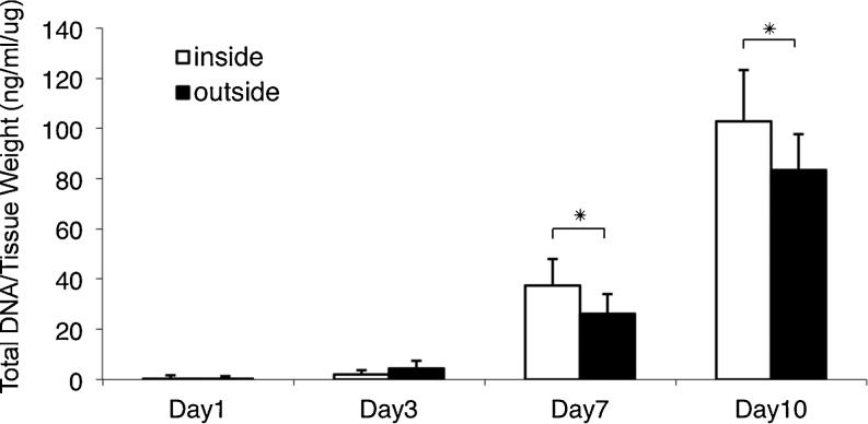
Methods: The synovium graft was placed within a complete tendon laceration, with either the visceral or parietal surface facing the proximal end of the lacerated tendon. The number of migrating cells was quantified by a cell migration assay. Qualitative immunohistochemistry and confocal laser microscopy were also used at day 10.
Results: Many labeled synovial cells were observed within the tendon to which the visceral surface of the synovium graft was facing. Migrated cells were also observed on the parietal side, but there were fewer cells compared to visceral surface cells. Migrating cells all expressed α-smooth muscle actin.
Conclusion: We found that graft orientation affected cell migration. Whether this finding has clinical significance awaits in vivo study.
Keywords: Cell migration; Synovial cells; Synovium graft; Tendon healing.
Figures


Similar articles
-
A histological observation on the flexor tendon healing within intact sheath.J Tongji Med Univ. 1991;11(3):169-73. doi: 10.1007/BF02888130. J Tongji Med Univ. 1991. PMID: 1784047
-
Synovial sheath cell migratory response to flexor tendon injury: an experimental study in rats.J Hand Surg Am. 2003 Nov;28(6):987-93. doi: 10.1016/s0363-5023(03)00380-0. J Hand Surg Am. 2003. PMID: 14642515
-
Histology and ultrastructure of the human flexor tendon sheath.J Hand Surg Am. 1987 Jan;12(1):25-9. doi: 10.1016/s0363-5023(87)80155-7. J Hand Surg Am. 1987. PMID: 3805639
-
Cellular architecture of the synovium in the tendon sheath of horses: an immunohistochemical and scanning electron microscopic study.Jpn J Vet Res. 2002 Nov;50(2-3):125-39. Jpn J Vet Res. 2002. PMID: 12619304
-
Stem cell therapies in tendon-bone healing.World J Stem Cells. 2021 Jul 26;13(7):753-775. doi: 10.4252/wjsc.v13.i7.753. World J Stem Cells. 2021. PMID: 34367476 Free PMC article. Review.
Cited by
-
Evaluation of the Effects of Synovial Multipotent Cells on Deep Digital Flexor Tendon Repair in a Large Animal Model of Intra-Synovial Tendinopathy.J Orthop Res. 2020 Jan;38(1):128-138. doi: 10.1002/jor.24423. Epub 2019 Aug 16. J Orthop Res. 2020. PMID: 31329308 Free PMC article.
References
-
- Amadio PC, Hunter JM, Jaeger SH, et al. The effect of vincular injury on the results of flexor tendon surgery in zone 2. J Hand Surg Am. 1985;10:626–632. - PubMed
-
- Beredjiklian PK. Biologic aspects of flexor tendon laceration and repair. J Bone Joint Surg Am. 2003;85-A:539–550. - PubMed
-
- Brockis JG. The blood supply of the flexor and extensor tendons of the fingers in man. J Bone Joint Surg Br. 1953;35-B:131–138. - PubMed
Publication types
Grants and funding
LinkOut - more resources
Full Text Sources
