. 2013 Nov 14:5:73.
doi: 10.3389/fnagi.2013.00073. eCollection 2013.

Age differences in the intrinsic functional connectivity of default network subsystems

Affiliations

Age differences in the intrinsic functional connectivity of default network subsystems

Karen L Campbell et al. Front Aging Neurosci. .

Abstract

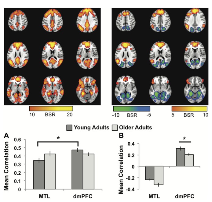
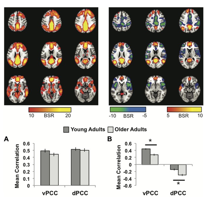
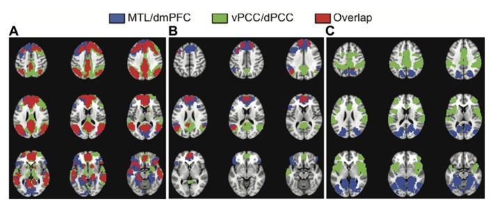
Recent work suggests that the default mode network (DMN) includes two core regions, the ventromedial prefrontal cortex and posterior cingulate cortex (PCC), and several unique subsystems that are functionally distinct. These include a medial temporal lobe (MTL) subsystem, active during remembering and future projection, and a dorsomedial prefrontal cortex (dmPFC) subsystem, active during self-reference. The PCC has been further subdivided into ventral (vPCC) and dorsal (dPCC) regions that are more strongly connected with the DMN and cognitive control networks, respectively. The goal of this study was to examine age differences in resting state functional connectivity within these subsystems. After applying a rigorous procedure to reduce the effects of head motion, we used a multivariate technique to identify both common and unique patterns of functional connectivity in the MTL vs. the dmPFC, and in vPCC vs. dPCC. All four areas had robust functional connectivity with other DMN regions, and each also showed distinct connectivity patterns in both age groups. Young and older adults had equivalent functional connectivity in the MTL subsystem. Older adults showed weaker connectivity in the vPCC and dmPFC subsystems, particularly with other DMN areas, but stronger connectivity than younger adults in the dPCC subsystem, which included areas involved in cognitive control. Our data provide evidence for distinct subsystems involving DMN nodes, which are maintained with age. Nevertheless, there are age differences in the strength of functional connectivity within these subsystems, supporting prior evidence that DMN connectivity is particularly vulnerable to age, whereas connectivity involving cognitive control regions is relatively maintained. These results suggest an age difference in the integrated activity among brain networks that can have implications for cognition in older adults.

Keywords: aging; default network; fMRI; functional connectivity; resting state; subsystems.


Figures

FIGURE 1
FIGURE 1
(A) The common network of regions positively correlated with MTL and dmPFC. (B) The regions uniquely correlated with the dmPFC (warm colors and positive correlations in the graph), or the MTL (cool colors and negative correlations in the graph). In both panels the graphs show the mean correlations (averaged across all 29 “blocks” in the resting run) between activity in each seed and the corresponding pattern of regions; i.e., the mean correlation between seed activity and the brain scores for young and older adults. In (B), warm-colored regions (i.e., regions with positive weights on the LV) are positively correlated with the dmPFC and cool-colored regions (negative weights) are positively correlated with the MTL. Error bars represent standard errors of the mean for the correlations. The color bars refer to the range of BSR values seen in the brain images (a BSR threshold of 10 was used). Asterisks indicate significant differences in functional connectivity (p <0.05, corrected). In this figure and subsequent ones, the images shown are from 48 mm above the AC-PC line to 16 mm below this line, in 8 mm increments.
FIGURE 2
FIGURE 2
(A) The common network of regions correlated with vPCC and dPCC. (B) The regions uniquely correlated with the vPCC (warm colors and positive correlations in the graph) or the dPCC (cool colors and negative correlations in the graph). In both panels the graphs show the mean correlations (averaged across all 29 “blocks” in the resting run) between activity in each seed and the corresponding pattern of regions; i.e., the mean correlation between seed activity and the brain scores for young and older adults. Error bars represent standard errors of the mean for the correlations. The color bars refer to the range of BSR values seen in the brain images (a BSR threshold of 5 was used). Asterisks indicate significant age group differences in functional connectivity (p <0.05, corrected).
FIGURE 3
FIGURE 3
The overlap between the common networks (LV1) (A), the dmPFC and vPCC subsystems (from LV2) (B), and the MTL and dPCC subsystems (from LV2) (C). There is considerable overlap between the common networks identified in the two analyses, including all major nodes of the DMN (A). There also is overlap in DMN regions between the dmPFC and vPCC subsystems, including superior frontal and angular gyri, in addition to the two seeds (B). There is essentially no overlap between the MTL and dPCC subsystems (C). A BSR threshold of 10 was used in (A) and a threshold of 5 was used in (B,C).

References

    1. Addis D., Wong A., Schacter D. (2007). Remembering the past and imagining the future: common and distinct neural substrates during even construction and elaboration. Neuropsychologia 45 1363–1377 10.1016/j.neuropsychologia.2006.10.016 - DOI - PMC - PubMed
    1. Allen E. A., Erhardt E. B., Damaraju E., Gruner W., Segall J. M., Silva R. F., et al. (2011). A baseline for the multivariate comparison of resting-state networks. Front. Syst. Neurosci. 5:2 10.3389/fnsys.2011.00002 - DOI - PMC - PubMed
    1. Andrews-Hanna J. R. (2012). The brain’s default network and its adaptive role in internal mentation. Neuroscientist 18 251–270 10.1177/1073858411403316 - DOI - PMC - PubMed
    1. Andrews-Hanna J. R., Reidler J. S., Sepulcre J., Poulin R., Buckner R. L. (2010). Functional-anatomic fractionation of the brain’s default network. Neuron 65 550–562 10.1016/j.neuron.2010.02.005 - DOI - PMC - PubMed
    1. Andrews-Hanna J. R., Snyder A. Z., Vincent J. L., Lustig C., Head D., Raichle M. E., et al. (2007). Disruption of large-scale brain systems in advanced aging. Neuron 56 924–935 10.1016/j.neuron.2007.10.038 - DOI - PMC - PubMed

LinkOut - more resources