

Pitfalls of contrast-enhanced ultrasound (CEUS) in the diagnosis of splenic sarcoidosis
- PMID: 24294346
- PMCID: PMC3774908
- DOI: 10.1007/s40477-013-0013-1
Pitfalls of contrast-enhanced ultrasound (CEUS) in the diagnosis of splenic sarcoidosis
Abstract
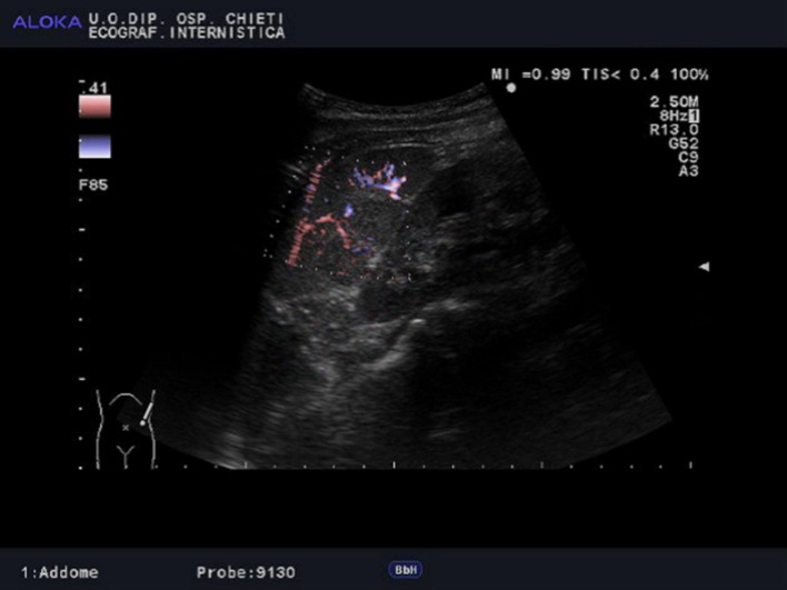
By observing the real-time behavior of focal liver lesions at three vascular phases (arterial, portal-venous, and late), contrast-enhanced ultrasound (CEUS) has been successfully applied to differentiate benign from malignant hepatic nodules. In recent years, numerous studies highlighted the usefulness of CEUS also for other applications such as abdominal trauma, renal, pancreatic, thyroid, and inflammatory bowel diseases, supporting its role even in differentiating benign from malignant splenic nodules. Therefore, the European Federation of Societies for Ultrasound in Medicine and Biology (EFSUMB) recently updated the guidelines for the use of ultrasound contrast agents in clinical practice, pointing out the indication to characterize splenic parenchymal inhomogeneity or suspected lesions found on conventional ultrasound (BUS). We describe the case of a patient with a history of colon cancer and finding, at BUS and CEUS, of hypoechoic lesions with a highly suggestive pattern for metastases, subsequently histologically proved to be splenic localizations of a benign and multisystemic granulomatous disease such as sarcoidosis. We therefore reviewed the current literature focusing on the role of CEUS in differentiating benign from malignant splenic lesions, emphasizing on the lack of data and numerical shortage of sarcoidosis derived-lesions in the available studies. We conclude that sarcoidosis remains a diagnosis of exclusion and new studies are needed before defining precise indications of CEUS in these patients.
L’ecografia con mezzo di contrasto (CEUS) è una metodica diagnostica minimamente invasiva ed ampiamente utilizzata, negli ultimi decenni, per differenziare le lesioni epatiche benigne dalle maligne, valutandone l’aspetto contrastografico nelle tre fasi vascolari (arteriosa, portale-venosa e tardiva). Negli ultimi anni, numerosi studi hanno messo in evidenza ulteriori indicazioni della CEUS, tra cui il trauma addominale, le lesioni focali renali, pancreatiche, tiroidee e le malattie infiammatorie intestinali, supportandone un ruolo anche nella differenziazione tra lesioni spleniche benigne e maligne. Pertanto, la Federazione Europea delle Società di Ultrasonologia in Medicina e Biologia (EFSUMB), ha recentemente aggiornato le linee guida per l’utilizzo dell’ecografia con mezzo di contrasto nella pratica clinica, sottolineando, tra le varie indicazioni, quella di caratterizzare le lesioni spleniche o disomogeneità parenchimali sospette, riscontrate incidentalmente all’esame ecografico convenzionale. Nel seguente articolo descriviamo il caso di una paziente con anamnesi per carcinoma del colon e riscontro, all’esame ecografico convenzionale, di lesioni ipoecogene di non univoca interpetazione e caratterizzate, alla CEUS, da un pattern contrastografico altamente suggestivo per metastasi. Tali lesioni si sono poi rivelate essere, all’esame istologico, localizzazioni di una malattia benigna, multisistemica e granulomatosa quale la sarcoidosi. Abbiamo pertanto effettuato una revisione approfondita della letteratura focalizzando sul ruolo della CEUS nel differenziare le lesioni spleniche benigne dalle maligne, enfatizzando sulla carenza di studi riguardanti la caratterizzazione, attraverso la CEUS, delle lesioni derivate dalla sarcoidosi. Tale malattia rimane, pertanto, una diagnosi di esclusione e sono necessari ulteriori studi prima di definire precise indicazioni della CEUS nei pazienti affetti.
Keywords: CEUS; Guidelines; Sarcoidosis; Spleen.
Figures


Similar articles
-
Value of Contrast-Enhanced Ultrasound in the Differential Diagnosis of Focal Splenic Lesions.Cancer Manag Res. 2021 Apr 1;13:2947-2958. doi: 10.2147/CMAR.S300601. eCollection 2021. Cancer Manag Res. 2021. PMID: 33833578 Free PMC article.
-
Incidentally detected splenic lesions in ultrasound: does contrast-enhanced ultrasonography improve the differentiation of benign hemangioma/hamartoma from malignant lesions?Ultraschall Med. 2011 Dec;32(6):582-92. doi: 10.1055/s-0031-1282034. Epub 2011 Dec 9. Ultraschall Med. 2011. PMID: 22161555
-
Contrast-enhanced ultrasound (CEUS): applications from the kidneys to the bladder.Abdom Radiol (NY). 2024 Nov;49(11):4092-4112. doi: 10.1007/s00261-024-04388-4. Epub 2024 Jun 17. Abdom Radiol (NY). 2024. PMID: 38884782 Review.
-
Clinical Applications of Contrast-Enhanced Ultrasound in the Pediatric Work-Up of Focal Liver Lesions and Blunt Abdominal Trauma: A Systematic Review.Ultrasound Int Open. 2017 Feb;3(1):E2-E7. doi: 10.1055/s-0042-124502. Ultrasound Int Open. 2017. PMID: 28255580 Free PMC article. Review.
-
Clinical use of contrast-enhanced ultrasound beyond the liver: a focus on renal, splenic, and pancreatic applications.Ultrasonography. 2019 Oct;38(4):278-288. doi: 10.14366/usg.18061. Epub 2018 Dec 30. Ultrasonography. 2019. PMID: 31230431 Free PMC article.
Cited by
-
CT Findings in Pulmonary and Abdominal Sarcoidosis. Implications for Diagnosis and Classification.J Clin Med. 2020 Sep 20;9(9):3028. doi: 10.3390/jcm9093028. J Clin Med. 2020. PMID: 32962242 Free PMC article. Review.
-
Hepatosplenic sarcoidosis: contrast-enhanced ultrasound findings and implications for clinical practice.Biomed Res Int. 2014;2014:926203. doi: 10.1155/2014/926203. Epub 2014 Aug 18. Biomed Res Int. 2014. PMID: 25215299 Free PMC article. Review.
-
Clinical Features, Histopathology and Differential Diagnosis of Sarcoidosis.Cells. 2021 Dec 26;11(1):59. doi: 10.3390/cells11010059. Cells. 2021. PMID: 35011621 Free PMC article. Review.
-
Hepatic artery resistive index as surrogate marker for fibrosis progression in NAFLD patients: A clinical perspective.Int J Immunopathol Pharmacol. 2018 Jan-Dec;32:2058738418781373. doi: 10.1177/2058738418781373. Int J Immunopathol Pharmacol. 2018. PMID: 29873275 Free PMC article.
-
Ultrasound imaging of abdominal sarcoidosis: State of the art.World J Clin Cases. 2019 Apr 6;7(7):809-818. doi: 10.12998/wjcc.v7.i7.809. World J Clin Cases. 2019. PMID: 31024952 Free PMC article. Review.
References
-
- Tana C, Tana M, Mezzetti A, Schiavone C. Sarcoidosis: old certainties and new perspectives. Ital J Med. 2012;6(3):186–194. doi: 10.1016/j.itjm.2012.05.002. - DOI
-
- Iannuzzi MC, Fontana JR (2011) Sarcoidosis: clinical presentation, immunopathogenesis, and therapeutics. JAMA 305(4):391–399 - PubMed
-
- Erdal BS, Clymer BD, Yildiz VO, Julian MW, Crouser ED (2012) Unexpectedly high prevalence of sarcoidosis in a representative U.S. Metropolitan population. Respir Med 106(6):893–899 (Epub 2012 Mar 13) - PubMed
LinkOut - more resources
Full Text Sources
Other Literature Sources
