Case Reports
. 2012 Dec;5(4):243-52.
doi: 10.1055/s-0032-1329542. Epub 2012 Nov 5.

Investigation of severe craniomaxillofacial battle injuries sustained by u.s. Service members: a case series

Affiliations
Case Reports

Investigation of severe craniomaxillofacial battle injuries sustained by u.s. Service members: a case series

Pamela R Brown Baer et al. Craniomaxillofac Trauma Reconstr. 2012 Dec.

Abstract

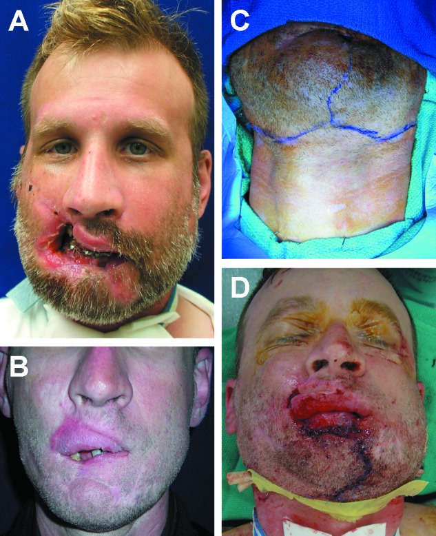
This case series describes craniomaxillofacial battle injuries, currently available surgical techniques, and the compromised outcomes of four service members who sustained severe craniomaxillofacial battle injuries in Iraq or Afghanistan. Demographic information, diagnostic evaluation, surgical procedures, and outcomes were collected and detailed with a follow-up of over 2 years. Reconstructive efforts with advanced, multidisciplinary, and multiple revision procedures were indicated; the full scope of conventional surgical options and resources were utilized. Patients experienced surgical complications, including postoperative wound dehiscence, infection, flap failure, inadequate mandibular healing, and failure of fixation. These complications required multiple revisions and salvage interventions. In addition, facial burns complicated reconstructive efforts by delaying treatment, decreasing surgical options, and increasing procedural numbers. All patients, despite multiple surgeries, continue to have functional and aesthetic deficits as a result of their injuries. Currently, no conventional treatments are available to satisfactorily reconstruct the face severely ravaged by explosive devices to an acceptable level, much less to natural form and function.

Keywords: craniomaxillofacial battle injuries; explosive injuries; reconstruction; surgical complications; surgical outcomes.


Figures

Figure 1
Figure 1
(A) One week post–explosive injury upon arrival to Brooke Army Medical Center. (B) Missing lower lip and chin apparent after release of primary lip repair. (C and D) Residual ramus stumps protruding through floor of mouth, bilaterally. (Images are courtesy of Colonel Robert Hale.)
Figure 2
Figure 2
(A and B) Titanium reconstruction plate to replace avulsed dentate mandible. Remaining lip and chin soft tissue closed over plate. (Images are courtesy of Colonel Robert Hale.)
Figure 3
Figure 3
Postoperative results after second osteofasciocutaneous fibula flap. (A) Microstomia and limited range of motion. (B) Lack of lower lip projection. (C) Skin paddle from free fibula flap replaces lower lip and chin. (Images are courtesy of Colonel Robert Hale.)
Figure 4
Figure 4
(A) Avulsion of two-thirds of the perioral structures after explosive injury. (B) Le Fort II fracture and comminuted mandibular body and rami from perforating shrapnel. (Images are courtesy of Colonel Robert Hale.)
Figure 5
Figure 5
(A and B) Primary fracture reconstruction; comminuted right condyle debridement and reconstructed with immediate costochondral graft. (Images are courtesy of Colonel Robert Hale.)
Figure 6
Figure 6
(A) Perioral deformity after skeletal repair. (B) Resultant deformity after bilateral cheek advancement flaps and reconstructive advancement of lower lip and a buccal mucosa advancement flap to reconstruct maxillary lip. (C) Incision markings for bilateral cervicofacial advancement/rotational flaps. (D) After cervicofacial flaps and anteriorly based ventral tongue flap to reconstruct lower lip. (Images are courtesy of Colonel Robert Hale.)
Figure 7
Figure 7
(A) Microstomia and dental condition after lip reconstruction. (B and C) Result after nine surgeries to reconstruct mandible fractures and perioral soft tissue avulsion. Reconstructed lips show microstomia, flaccidity, and lack of projection. (Images are courtesy of Colonel Robert Hale.)
Figure 8
Figure 8
(A) Second- and third-degree facial burns and perforating wounds to right orbit, nasofrontal area, and right check after explosive injury. (B) Comminuted frontal sinus, naso-orbital-ethmoid, and Le Fort II fractures. (Images are courtesy of Colonel Robert Hale.)
Figure 9
Figure 9
(A) Burned skin debrided and closed with meshed split thickness skin grafts. (B) Nine months postinjury face resurfaced with transpositional, expanded supraclavicular flaps. (Images are courtesy of Colonel Robert Hale.)
Figure 10
Figure 10
(A and B) Result after 26 operations. Naso-orbital skeletal deformity and nasal soft tissue persists. Poor skin quality due to facial burns was main limiting factor. (Images are courtesy of Colonel Robert Hale.)
Figure 11
Figure 11
(A and B) Second- and third-degree facial burns after injury by an explosive device. Eyes are protected with supratarsal release and full-thickness skin grafts and eyelids sutured together to mitigate ectropion. Plastic lip retractor mitigates microstomia as burned skin contracts during healing. (Images are courtesy of Colonel Robert Hale.)
Figure 12
Figure 12
(A and B) Computed tomography scans show comminuted anterior mandibular and bilateral condylar fractures. Fractures were stabilized with MMF for 12 weeks. (Images are courtesy of Colonel Robert Hale.) MMF, maxillomandibular fixation.
Figure 13
Figure 13
Nine months after injury. (A and B) Mandibular ankylosis and malunion are apparent. (C, D, and E) Severe contractures, hypertrophic scars, ectropion, lip retraction, microstomia, and limited range of motion are directly related to burns and mandibular fractures. (Images are courtesy of Colonel Robert Hale.)
Figure 14
Figure 14
(A and B) Result following 27 facial procedures. Patient still suffers from microstomia, decreased range of mandibular motion, ectropion of lower eyelids and nasal dysfunction, facial scarring, and deformities. (Images are courtesy of Colonel Robert Hale.)

References

    1. Lew T A, Walker J A, Wenke J C, Blackbourne L H, Hale R G. Characterization of craniomaxillofacial battle injuries sustained by United States service members in the current conflicts of Iraq and Afghanistan. J Oral Maxillofac Surg. 2010;68:3–7. - PubMed
    1. Owens B D, Kragh J F Jr, Wenke J C, Macaitis J, Wade C E, Holcomb J B. Combat wounds in operation Iraqi Freedom and operation Enduring Freedom. J Trauma. 2008;64:295–299. - PubMed
    1. Masini B D, Waterman S M, Wenke J C, Owens B D, Hsu J R, Ficke J R. Resource utilization and disability outcome assessment of combat casualties from Operation Iraqi Freedom and Operation Enduring Freedom. J Orthop Trauma. 2009;23:261–266. - PubMed
    1. Kauvar D S, Wolf S E, Wade C E, Cancio L C, Renz E M, Holcomb J B. Burns sustained in combat explosions in Operations Iraqi and Enduring Freedom (OIF/OEF explosion burns) Burns. 2006;32:853–857. - PubMed
    1. Motamedi M H. Primary treatment of penetrating injuries to the face. J Oral Maxillofac Surg. 2007;65:1215–1218. - PubMed

Publication types

LinkOut - more resources