Abnormality of the Foramen Spinosum due to a Variation in the Trajectory of the Middle Meningeal Artery: A Case Report in Human
- PMID: 24294564
- PMCID: PMC3836884
- DOI: 10.1055/s-0033-1347901
Abnormality of the Foramen Spinosum due to a Variation in the Trajectory of the Middle Meningeal Artery: A Case Report in Human
Abstract
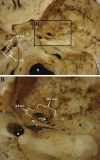
Originating from the maxillary artery, the middle meningeal artery (MMA) is predominantly periosteal irrigating the bone and dura mater. It enters the floor of the middle cranial fossa through the foramen spinosum, travels laterally through a middle fossa bony ridge, and curves over the previous upper-greater wing of the sphenoid, where it in a variable point is divided into frontal and parietal branches. The complex sequence of the MMA development gives many opportunities for variant anatomy. In a Caucasian cadaver skull of an approximately 35-year-old individual belonging to the didactical collection of the Laboratory of Human Anatomy at the University of Santa Cruz do Sul, Brazil, it was noted that the right foramen spinosum has an abnormal shape. In this report, we discuss an abnormality of the foramen spinosum due to a variation in the trajectory of the MMA. Thus, the present study shall be important for health sciences and those who have some interest in pathologies associated with the MMA.
Keywords: anatomic variation; foramen ovale; foramen spinosum; middle meningeal artery.
Figures

Similar articles
-
Bony Tunnel Formation Associated with the Distal Segment of the Frontal Branch of the Middle Meningeal Artery.J Neurol Surg B Skull Base. 2019 Oct;80(5):480-483. doi: 10.1055/s-0038-1676353. Epub 2018 Dec 3. J Neurol Surg B Skull Base. 2019. PMID: 31534889 Free PMC article.
-
Absence and Duplicate Foramen Spinosum in the Same Patient: An Extremely Rare Variation.J Craniofac Surg. 2024 Jul-Aug 01;35(5):e474-e476. doi: 10.1097/SCS.0000000000010353. Epub 2024 May 30. J Craniofac Surg. 2024. PMID: 38814095
-
Absence of foramen spinosum and abnormal middle meningeal artery in cranial series.Anthropol Anz. 2012 Jul;69(3):351-66. doi: 10.1127/0003-5548/2012/0165. Anthropol Anz. 2012. PMID: 22928356
-
Unilateral absence of foramen spinosum with bilateral ophthalmic origin of the middle meningeal artery: case report and review of the literature.Folia Morphol (Warsz). 2014 Feb;73(1):87-91. doi: 10.5603/FM.2014.0013. Folia Morphol (Warsz). 2014. PMID: 24590529 Review.
-
Skull base fracture involving the foramen spinosum - an indirect sign of middle meningeal artery lesion: case report and literature review.Turk Neurosurg. 2015;25(2):317-9. doi: 10.5137/1019-5149.JTN.7875-13.1. Turk Neurosurg. 2015. PMID: 26014021 Review.
Cited by
-
Proximity of the middle meningeal artery and maxillary artery to the mandibular head and mandibular neck as revealed by three-dimensional time-of-flight magnetic resonance angiography.Oral Maxillofac Surg. 2022 Mar;26(1):139-146. doi: 10.1007/s10006-021-00960-0. Epub 2021 May 23. Oral Maxillofac Surg. 2022. PMID: 34024006 Free PMC article.
-
Topographic and Morphometric Study of the Foramen Spinosum of the Skull and Its Clinical Correlation.Medicina (Kaunas). 2022 Nov 28;58(12):1740. doi: 10.3390/medicina58121740. Medicina (Kaunas). 2022. PMID: 36556942 Free PMC article.
References
-
- Kresimir Lukic I, Gluncic V, Marusic A. Extracranial branches of the middle meningeal artery. Clin Anat. 2001;14(4):292–294. - PubMed
-
- Kuruvilla A, Aguwa A N, Lee A W, Xavier A R. Anomalous origin of the middle meningeal artery from the posterior inferior cerebellar artery. J Neuroimaging. 2011;21(3):269–272. - PubMed
LinkOut - more resources
Full Text Sources
Other Literature Sources

