

Epiretinal membranes in patients with uveitis: morphological and functional analysis with spectral domain optical coherence tomography
- PMID: 24294602
- PMCID: PMC3835473
- DOI: 10.1155/2013/284821
Epiretinal membranes in patients with uveitis: morphological and functional analysis with spectral domain optical coherence tomography
Abstract
Purpose: To correlate the uveitic epiretinal membrane (ERM) features using spectral-domain optical coherence tomography (SD-OCT) with visual acuity (VA).
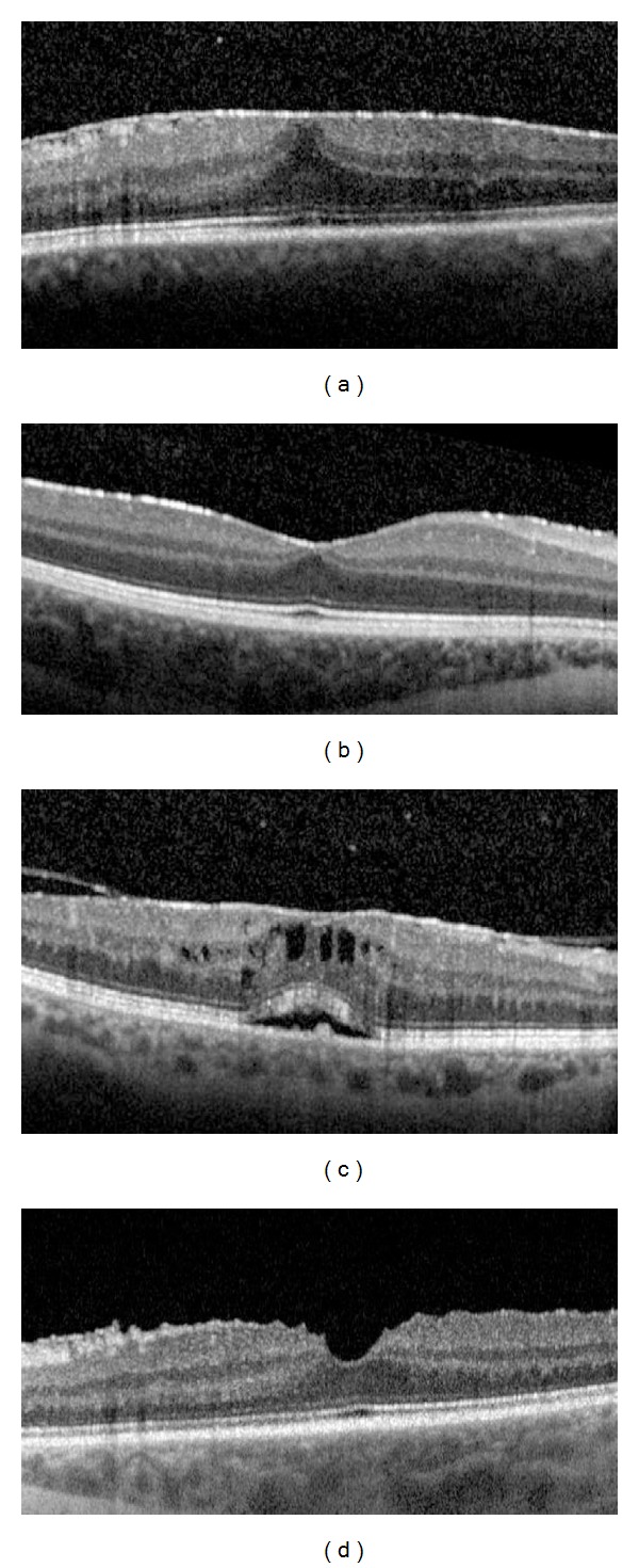
Methods: Forty-one eyes of 32 patients were included in this retrospective study. SD-OCT was performed in all patients and data were collected at the time of ERM diagnosis and at the final visit. Both best corrected visual acuity (BCVA) and ERM thickness were correlated with the morphological and clinical features.
Results: Final BCVA was positively correlated with male sex (P = 0.0055) and the focal pattern of ERM attachment (P = 0.031) and negatively correlated with IS/OS photoreceptor junction disruption (P = 0.042). BVCA change showed a positive correlation with the age of ERM onset (P = 0.056) but a negative correlation with IS/OS photoreceptor disruption at the ERM diagnosis (P = 0.029) and the increase of central subfield thickness (CST) (P = 0.95). Final ERM thickness correlated with the duration of uveitis (P = 0.0023) and the duration of ERM (P = 1.15 e-05). During the follow-up, ERM thickening correlated with male sex (P = 0.042), posterior uveitis (P = 0.036), uveitis duration (P = 0.026), and broad attachment pattern (P = 0.052).
Conclusions: In the uveitic ERM, VA negatively correlates with IS/OS photoreceptor junction disruption and the increase of CST. ERM thickness is influenced by longer duration of both uveitis and ERM.
Figures


Similar articles
-
Correlation between visual acuity changes and optical coherence tomography morphological findings in idiopathic epiretinal membranes.Graefes Arch Clin Exp Ophthalmol. 2016 Mar;254(3):437-44. doi: 10.1007/s00417-015-3069-0. Epub 2015 May 29. Graefes Arch Clin Exp Ophthalmol. 2016. PMID: 26016811
-
Morphometric spectral-domain optical coherence tomography features of epiretinal membrane correlate with visual acuity in patients with uveitis.Am J Ophthalmol. 2012 Jul;154(1):78-86.e1. doi: 10.1016/j.ajo.2012.01.032. Epub 2012 Apr 26. Am J Ophthalmol. 2012. PMID: 22541930 Free PMC article.
-
Disruption of the photoreceptor inner segment/outer segment layer on spectral domain-optical coherence tomography is a predictor of poor visual acuity in patients with epiretinal membranes.Retina. 2010 May;30(5):713-8. doi: 10.1097/IAE.0b013e3181c596e3. Retina. 2010. PMID: 20038861
-
[Correlation between the morphology of the IS/OS junction and functional outcomes in patients with idiopathic epiretinal membrane].Nippon Ganka Gakkai Zasshi. 2012 Nov;116(11):1029-36. Nippon Ganka Gakkai Zasshi. 2012. PMID: 23316651 Review. Japanese.
-
THE EFFECT OF INTERNAL LIMITING MEMBRANE PEELING ON IDIOPATHIC EPIRETINAL MEMBRANE SURGERY, WITH A REVIEW OF THE LITERATURE.Retina. 2017 May;37(5):873-880. doi: 10.1097/IAE.0000000000001263. Retina. 2017. PMID: 27617536 Review.
Cited by
-
Epiretinal membrane: optical coherence tomography-based diagnosis and classification.Clin Ophthalmol. 2016 Mar 29;10:527-34. doi: 10.2147/OPTH.S97722. eCollection 2016. Clin Ophthalmol. 2016. PMID: 27099458 Free PMC article. Review.
-
Functional and Anatomical Outcomes of Pars Plana Vitrectomy for Epiretinal Membrane in Patients with Uveitis.Diagnostics (Basel). 2022 Dec 5;12(12):3044. doi: 10.3390/diagnostics12123044. Diagnostics (Basel). 2022. PMID: 36553051 Free PMC article.
-
Correlation between Morphological Characteristics of Macular Edema and Visual Acuity in Young Patients with Idiopathic Intermediate Uveitis.Medicina (Kaunas). 2023 Mar 8;59(3):529. doi: 10.3390/medicina59030529. Medicina (Kaunas). 2023. PMID: 36984530 Free PMC article.
-
Photoreceptor inner segment ellipsoid band integrity on spectral domain optical coherence tomography.Clin Ophthalmol. 2014 Dec 9;8:2507-22. doi: 10.2147/OPTH.S72132. eCollection 2014. Clin Ophthalmol. 2014. PMID: 25525329 Free PMC article. Review.
-
Correlation between visual acuity changes and optical coherence tomography morphological findings in idiopathic epiretinal membranes.Graefes Arch Clin Exp Ophthalmol. 2016 Mar;254(3):437-44. doi: 10.1007/s00417-015-3069-0. Epub 2015 May 29. Graefes Arch Clin Exp Ophthalmol. 2016. PMID: 26016811
References
-
- Charles S. Techniques and tools for dissection of epiretinal membranes. Graefe’s Archive for Clinical and Experimental Ophthalmology. 2003;241(5):347–352. - PubMed
-
- Smiddy WE, Maguire AM, Green WR, et al. Idiopathic epiretinal membranes: ultrastructural characteristics and clinicopathologic correlation. Retina. 2005;25(5):811–821. - PubMed
-
- Kampik A, Kenyon KR, Michels RG, Green WR, de la Cruz ZC. Epiretinal and vitreous membranes: comparative study of 56 cases. Retina. 2005;25(5):1445–1454. - PubMed
-
- Puliafito CA, Hee MR, Lin CP, et al. Imaging of macular diseases with optical coherence tomography. Ophthalmology. 1995;102(2):217–229. - PubMed
MeSH terms
LinkOut - more resources
Full Text Sources
Other Literature Sources
