Review
. 2014 May 10;20(14):2160-78.
doi: 10.1089/ars.2013.5731. Epub 2014 Feb 25.

Cellular biochemistry methods for investigating protein tyrosine phosphatases

Affiliations
Review

Cellular biochemistry methods for investigating protein tyrosine phosphatases

Stephanie M Stanford et al. Antioxid Redox Signal. .

Abstract

Significance: The protein tyrosine phosphatases (PTPs) are a family of proteins that play critical roles in cellular signaling and influence many aspects of human health and disease. Although a wealth of information has been collected about PTPs since their discovery, many questions regarding their regulation and function still remain.

Critical issues: Of particular importance are the elucidation of the biological substrates of individual PTPs and understanding of the chemical and biological basis for temporal and spatial resolution of PTP activity within a cell.

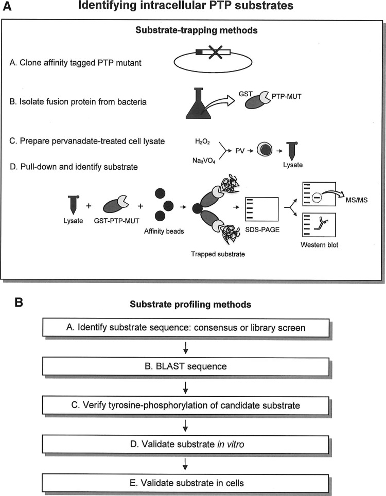
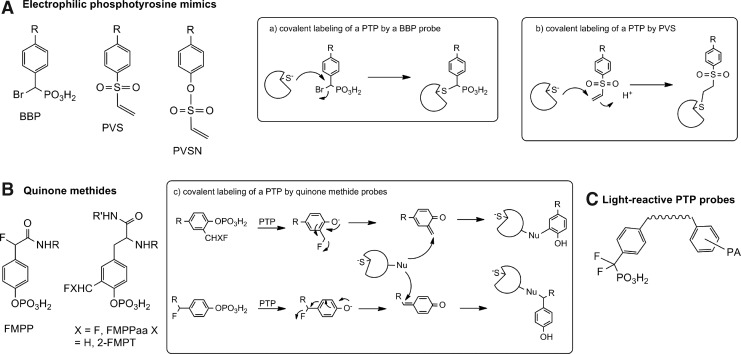
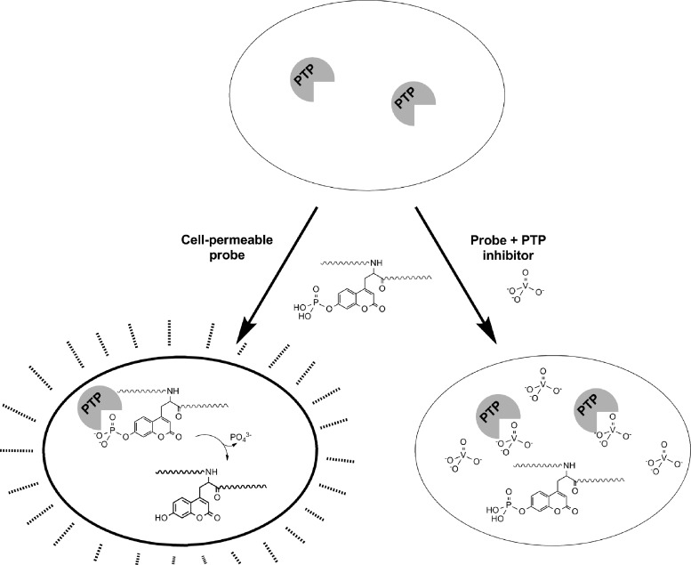
Recent advances: Drawing from recent advances in both biology and chemistry, innovative approaches have been developed to study the intracellular biochemistry and physiology of PTPs. We provide a summary of PTP-tailored techniques and approaches, emphasizing methodologies to study PTP activity within a cellular context. We first provide a discussion of methods for identifying PTP substrates, including substrate-trapping mutants and synthetic peptide libraries for substrate selectivity profiling. We next provide an overview of approaches for monitoring intracellular PTP activity, including a discussion of mechanistic-based probes, gel-based assays, substrates that can be used intracellularly, and assays tied to cell growth. Finally, we review approaches used for monitoring PTP oxidation, a key regulatory pathway for these enzymes, discussing the biotin switch method and variants of this approach, along with affinity trapping techniques and probes designed to detect PTP oxidation.

Future directions: Further development of approaches to investigate the intracellular PTP activity and functions will provide specific insight into their mechanisms of action and control of diverse signaling pathways.


Figures

<b>FIG. 1.</b>
FIG. 1.
Classification system of protein tyrosine phosphatases (PTPs) in the human genome. PTPs have been assigned to four classes based upon the amino acid sequence of their catalytic domain (3). Class I Cys-based PTPs evolved from a common ancestor and include the classical Tyr-specific transmembrane and nonreceptor PTPs and the various subclasses of dual-specificity PTPs (DUSPs). Class II contains the Cys-based low-molecular-weight PTP (LMPTP), which is structurally similar to bacterial arsenate reductases. Class III contains the Tyr/Thr-specific Cys-based CDC25 PTPs. Class IV contains the Eyes Absent PTPs, which utilize an Asp-based catalytic mechanism. Additional novel PTPs have been more recently discovered, the ubiquitin-associated and SH3 domain containing B (UBASH3B), which contains a histidine phosphatase domain (85), and suppressor of SUa7 gene 2 (SSU72), which has a catalytic domain structure similar to that of LMPTP (80, 142).
<b>FIG. 2.</b>
FIG. 2.
Methods to identify intracellular PTP substrates. Traditional approaches to identify PTP substrates have included (A) substrate trapping, in which phosphorylated substrates from pervanadate-stimulated cells are pulled down by recombinant PTP-trapping mutants and identified by mass spectrometry or western blotting, and (B) substrate profiling based upon amino acid sequence similarity to a previously identified protein or peptidic substrate.
<b>FIG. 3.</b>
FIG. 3.
Electrophilic, mechanism-based covalent inhibitors of PTP activity. (A) Bromobenzylphosphonates (BBP), phenylvinylsulfones (PVS), and phenyl vinylsulfonates (PVSN) are three categories of phosphotyrosine (pTyr) mimetic electrophilic moieties that can form a covalent bond with the catalytic Cys residue. (B) Quinone methides are substrate analogs that form a reactive electrophile upon PTP-mediated dephosphorylation. This electrophile can then covalently attach to a nucleophilic residue on the PTP or other adjacent biomolecule. (C) A pTyr mimetic phosphonate group can be used to localize a photoactive reagent near the enzyme.
<b>FIG. 4.</b>
FIG. 4.
Fluorogenic PTP substrates. Dephosphorylation of phosphorylated fluorescein and coumarin substrates results in a large increase in fluorescence and these types of substrates are commonly used to assay PTP activity. (A) Fluorescein diphosphate (FDP) can be caged to create an inert but photoactivatable prosubstrate for use in cells. (B) A ratiometric probe combining the FDP substrate with a fluorescence resonance energy transfer (FRET)-paired donor coumarin fluorophore. This substrate is also a prosubstrate that must be activated by cellular esterases. Once activated, in the absence of PTP activity, excitation of the coumarin fluorophore will result in coumarin emission. In the presence of PTP activity, the FDP moiety will be dephosphorylated and excitation of the coumarin fluorophore will result in FRET to the fluorescein and subsequent fluorescein emission. (C) A phosphorylated coumarin amino acid that can be incorporated into a cell-permeable peptide substrate and delivered to cells.
<b>FIG. 5.</b>
FIG. 5.
Cell-based assay for PTP inhibitors with fluorogenic pTyr mimic. Cells are incubated with a cell-permeable, fluorogenic pTyr mimic such as a phosphorylated coumaryl amino propionic acid (pCAP)-containing peptide. Once internalized, the substrate is exposed to intracellular PTPs, which dephosphorylate the substrate. Exposure of the cells to UV or violet light excites the dephosphorylated Tyr mimic, and fluorescence can be detected by fluorescence acquired cell sorting or microscopy. In the presence of cell-permeable PTP inhibitors, however, the substrate is not hydrolyzed and the cell fluorescence is attenuated.
<b>FIG. 6.</b>
FIG. 6.
Light-reactive covalent inhibitors of PTP activity. The phosphate groups of a quinone methide probe can be caged or masked by using photolabile esters. The unreactive caged probe can be delivered to cells and activated at the desired time.
<b>FIG. 7.</b>
FIG. 7.
Redox regulation of PTPs. PTPs can be regulated by oxidation. Exposure to biological agents, such as nitric oxide and hydrogen peroxide, leads to the formation of an S-nitrosocysteine or sulfenic acid. These adducts are reversible, and the S-nitrosocysteine can be converted into sulfenic acid by hydrolysis. Some PTPs form a stable, sulfenic acid adduct, which can be further oxidized, resulting in irreversible inactivation of the phosphatase by formation of a sulfinic or sulfonic acid species.
<b>FIG. 8.</b>
FIG. 8.
Monitoring the cellular redox state of the PTPs. Intracellular PTPs may be reversibly oxidized (represented as a sulfenic acid in this scheme) or irreversibly oxidized (represented as a sulfonic acid here). (A) The biotin switch method for identifying reversibly oxidized PTPs in cells sets the standard. Cells can be exposed to a specific stimulus and then lysed under anaerobic conditions. Any PTP with a reduced catalytic Cys residue can be alkylated with a reagent such as iodoacetic acid (IAA) (this also serves to alkylate any other free Cys residues in the lysate). Under the appropriate reducing conditions, disulfides, sulfenic acids, and S-nitrosocysteine residues can be reduced to reactivate reversibly oxidized PTPs. A thiol-reactive biotin reagent can be used to pull down the active PTPs. (B) In a variation of the previous procedure, a PTP of interest can be affinity trapped out of a lysate and the activity of that PTP monitored directly using a fluorogenic substrate. (C) The dimedone chemical probe can be used to identify PTPs that are specifically modified to form sulfenic acids in cells.

Similar articles

Cited by

References

    1. Agazie YM. and Hayman MJ. Development of an efficient “substrate-trapping” mutant of Src homology phosphotyrosine phosphatase 2 and identification of the epidermal growth factor receptor, Gab1, and three other proteins as target substrates. J Biol Chem 278: 13952–13958, 2003 - PubMed
    1. Almo SC, Bonanno JB, Sauder JM, Emtage S, Dilorenzo TP, Malashkevich V, et al. . Structural genomics of protein phosphatases. J Struct Funct Genomics 8: 121–140, 2007 - PMC - PubMed
    1. Alonso A, Sasin J, Bottini N, Friedberg I, Friedberg I, Osterman A, et al. . Protein tyrosine phosphatases in the human genome. Cell 117: 699–711, 2004 - PubMed
    1. Andersen JN, Mortensen OH, Peters GH, Drake PG, Iversen LF, Olsen OH, et al. . Structural and evolutionary relationships among protein tyrosine phosphatase domains. Mol Cell Biol 21: 7117–7136, 2001 - PMC - PubMed
    1. Baba H, Masuda Y, Sueyoshi N, and Kameshita I. In-gel phosphatase assay using non-denaturing two-dimensional electrophoresis. J Biochem 152: 557–563, 2012 - PubMed

Publication types

MeSH terms

Substances

LinkOut - more resources