Review
. 2014 Sep 1;21(7):1119-42.
doi: 10.1089/ars.2013.5486. Epub 2014 Feb 19.

Reactive oxygen and nitrogen species in steatotic hepatocytes: a molecular perspective on the pathophysiology of ischemia-reperfusion injury in the fatty liver

Affiliations
Review

Reactive oxygen and nitrogen species in steatotic hepatocytes: a molecular perspective on the pathophysiology of ischemia-reperfusion injury in the fatty liver

Megan J Reiniers et al. Antioxid Redox Signal. .

Abstract

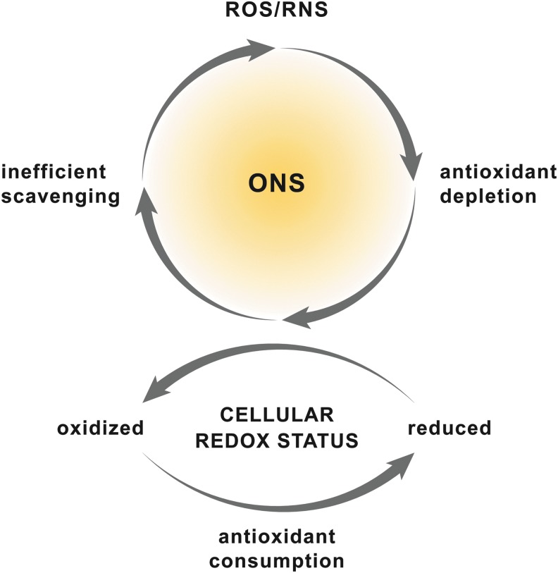
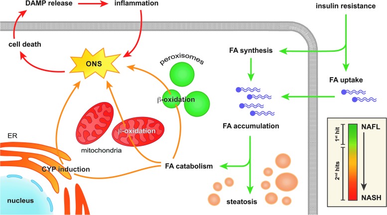
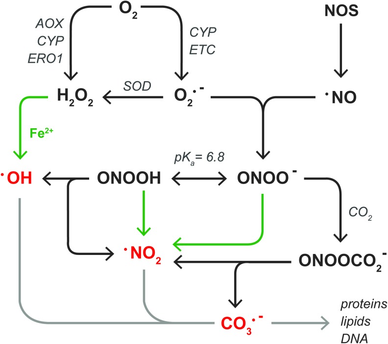
Significance: Hepatic ischemia-reperfusion (IR) injury results from the temporary deprivation of hepatic blood supply and is a common side effect of major liver surgery (i.e., transplantation or resection). IR injury, which in most severe cases culminates in acute liver failure, is particularly pronounced in livers that are affected by non-alcoholic fatty liver disease (NAFLD). In NAFLD, fat-laden hepatocytes are damaged by chronic oxidative/nitrosative stress (ONS), a state that is acutely exacerbated during IR, leading to extensive parenchymal damage.

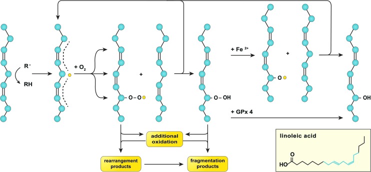
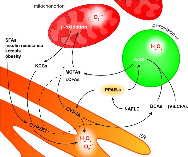
Recent advances: NAFLD triggers ONS via increased (extra)mitochondrial fatty acid oxidation and activation of the unfolded protein response. ONS is associated with widespread protein and lipid (per)oxidation, which reduces the hepatic antioxidative capacity and shifts the intracellular redox status toward an oxidized state. Moreover, activation of the transcription factor peroxisome proliferator-activated receptor α induces expression of mitochondrial uncoupling protein 2, resulting in depletion of cellular energy (ATP) reserves. The reduction in intracellular antioxidants and ATP in fatty livers consequently gives rise to severe ONS and necrotic cell death during IR.

Critical issues: Despite the fact that ONS mediates both NAFLD and IR injury, the interplay between the two conditions has never been described in detail. An integrative overview of the pathophysiology of NAFLD that renders steatotic hepatocytes more vulnerable to IR injury is therefore presented in the context of ONS.

Future directions: Effective methods should be devised to alleviate ONS and the consequences thereof in NAFLD before surgery in order to improve resilience of fatty livers to IR injury.


Figures

<b>FIG. 1.</b>
FIG. 1.
Etiology of ONS. ONS is the result of a disrupted balance between intracellular levels of ROS/RNS and antioxidants. The production of large amounts of ROS/RNS consumes cellular antioxidants, which leads to inefficient ROS/RNS scavenging and hence ONS (upper panel). Simultaneously, the cellular redox status shifts toward a more oxidized state. Alternatively, disruption of the cellular redox status can induce ONS because of the consequent consumption and depletion of cellular antioxidants (bottom panel). ONS, oxidative/nitrosative stress; RNS, reactive nitrogen species; ROS, reactive oxygen species. To see this illustration in color, the reader is referred to the web version of this article at www.liebertpub.com/ars
<b>FIG. 2.</b>
FIG. 2.
Pathogenesis of NAFLD. Hepatic insulin resistance increases the uptake as well as the synthesis of FAs by hepatocytes, which results in the accumulation of FAs that are stored as triglycerides and the development of steatosis (green arrows). In addition, FA-catabolizing pathways become hyperactivated to dispose of the excess in FAs (orange arrows) through increased mitochondrial and peroxisomal β-oxidation as well as the induction of CYP enzymes in the ER. Since all of these FA catabolizing pathways produce oxidants, ONS ensues, which eventually induces cell death and the release of DAMPs. Thereafter, DAMPs incite an immune response that further aggravates the ONS and consequently induces cell death, creating a positive feedback loop that eventually leads to pervasive parenchymal inflammation (red arrows). The inset shows how the phenomena mentioned earlier (i.e., FA accumulation, FA catabolism, and inflammation) relate to the pathological stage of NAFLD, ranging from NAFL to NASH (right of scale bar), and the putative two-hit theory (left of scale bar). The scale bar is color matched to the figure arrows. CYP, cytochrome P450; DAMP, damage-associated molecular pattern; ER, endoplasmic reticulum; FA, fatty acid; NAFL, non-alcoholic fatty liver; NAFLD, non-alcoholic fatty liver disease; NASH, non-alcoholic steatohepatitis. To see this illustration in color, the reader is referred to the web version of this article at www.liebertpub.com/ars
<b>FIG. 3.</b>
FIG. 3.
ROS and RNS relevant to NAFLD and hepatic IR. Superoxide anion (O2•−) is formed in the one-electron reduction of O2 by CYP enzymes or the ETC. Thereafter, different SODs catalyze the dismutation of O2•− into hydrogen peroxide (H2O2). In addition, H2O2 is formed in the two-electron reduction of O2 by AOX, CYP enzymes, or ERO1. H2O2 can react with transition metals such as ferrous iron (Fe2+, green arrows) to form hydroxyl radical (•OH). Alternatively, O2•− reacts with NOS-derived nitric oxide (•NO) to form peroxynitrite anion (ONOO), which exists in equilibrium with its conjugate acid peroxynitrous acid (ONOOH, pKa=6.8). Both forms of peroxynitrite can react with Fe2+ to generate nitrogen dioxide (•NO2). •NO2 is also formed during the homolytic fission of ONOOH, along with •OH, as well as in the reaction between ONOO- and CO2, also yielding carbonate radical anion (CO3•−). The free radicals •OH, •NO2, and CO3•− are indicated in red to emphasize their high reactivity, which enables them to irreversibly alter the chemical structure of biomolecules (light gray arrow) as described in section “Molecular targets of ROS/RNS in NAFLD and IR.” AOX, fatty acyl-CoA oxidase; ERO1, ER oxidoreductin 1; ETC, electron transport chain; IR, ischemia-reperfusion; NOS, nitric oxide synthase; SOD, superoxide dismutase. To see this illustration in color, the reader is referred to the web version of this article at www.liebertpub.com/ars
<b>FIG. 4.</b>
FIG. 4.
Lipid peroxidation. Lipid peroxidation is initiated by hydrogen abstraction, mostly from hydrocarbons that are flanked by two alkenes (depicted on the left), that is, those on the aliphatic chains of PUFAs such as linoleic acid (inset, in which the structure highlighted in blue corresponds to the aliphatic structure in the figure). The formed carbon-centered lipid radical (L•, yellow dot) has the ability to relocate three carbon atoms away from the abstraction site (dashed line), where it swiftly reacts with oxygen to form a lipid peroxyl radical (L–OO•). Subsequently, L–OO• can undergo intramolecular modification to form rearrangement products, additional oxidation, or react with a proximal PUFA to generate a lipid hydroperoxide (L–OOH) as well as a new L• (top arrow). L–OOH can undergo additional oxidation or dissociate into fragmentation products. Alternatively, L–OOH can react with GPx 4 to form a lipid hydroxide (L–OH) or undergo ferrous iron (Fe2+)-catalyzed oxidation to form a lipid alkoxyl radical (L–O•), the latter of which has the ability to oxidize another PUFA and generate a new L• (top arrow) as well as a L–OH. All these events lead to membrane destabilization as a result of lipid packing defects, which may have detrimental consequences on cell function and viability. GPx 4, glutathione peroxidase 4; PUFA, polyunsaturated fatty acid. To see this illustration in color, the reader is referred to the web version of this article at www.liebertpub.com/ars
<b>FIG. 5.</b>
FIG. 5.
Free radical scavenging in the lipid compartment. Initiators of lipid peroxidation such as lipid peroxyl radicals (L–OO•) and nitrogen dioxide (•NO2) are scavenged by α-tocopherol during which an α-tocopherol radical (α-tocopherol•) is formed. The α-tocopherol radical is subsequently reduced by ascorbic acid (asc. acid) or ubiquinol (UQH2, depicted on the left), resulting in the formation of an ascorbic acid (asc. acid•) or ubisemiquinone radical (UQH•), respectively. Alternatively, UQH• is formed during the direct reduction of L–OO• or •NO2 by UQH2 (depicted on the right). UQH• reacts with O2 to form UQ as well as superoxide anion (O2•−). UQ can be regenerated into UQH2 via reduction by the mitochondrial ETC, but UQH2 is also synthesized endogenously from acetyl CoA via the MP (upper right corner). In addition, UQ can undergo (auto-)oxidation (ox.) into inactive metabolites (lower right corner). MP, mevalonate pathway; UQ/UQH2, ubiquinone/ubiquinol, also referred to as coenzyme Q10. To see this illustration in color, the reader is referred to the web version of this article at www.liebertpub.com/ars
<b>FIG. 6.</b>
FIG. 6.
Mechanisms of protein oxidation. Six mechanisms of protein oxidation are depicted (yellow bars and numbers, top and center). The species of ROS/RNS involved in each mechanism are listed above. The top scheme represents the protein backbone with the individual amino acids (see in-figure legend) in which the polygonal/circular forms represent aromatic structures on the respective amino acid residues. The sites of oxidation by ROS/RNS are indicated with orange shading. The bottom scheme shows the structural changes after oxidation, also higlighted in orange. Details on the reaction mechanisms are provided in the text. To see this illustration in color, the reader is referred to the web version of this article at www.liebertpub.com/ars
<b>FIG. 7.</b>
FIG. 7.
TNF-α signaling in hepatocytes. After TNF-α binding to TNFR1, TRADD, TRAF2, and RIP1 are assembled at the cytosolic domain of TNFR1 to form complex 1 (top center). Complex 1 subsequently initiates two branches of the trinomial death signaling cascade via the phosphorylation of JNK (pathway A) as well as the accumulation of ceramide and the subsequent increase in GGD3 synthesis (pathway B). Both routes increase mitochondrial O2•− formation (bottom panel), although the exact mechanism of JNK-mediated O2•− formation remains elusive (dashed line). The formation of derivative ROS/RNS from O2•− and corollary activation of cytochrome c peroxidase activity subsequently leads to the dissociation of cytochrome c from the IMM (bottom panel), a prerequisite for apoptosis. In addition, sustained JNK-dependent ROS/RNS formation leads to MPT and necrosis. Simultaneously, complex 1 triggers survival signaling via activation of the IKK complex (pathway D, top right), which phosphorylates IκB-α. This enables the dissociation of IκB-α from NF-κB and its translocation to the nucleus to initiate the transcription of anti-apoptotic and antioxidative genes. At a later time point, TRADD and RIP1 dissociate from complex 1 and associate with FADD, procaspase 8, and RIP3 to form complex 2 (upper left corner). Complex 2 mediates the third branch of the death signaling cascade (pathway C) by activating caspase 8, which subsequently truncates Bid into tBid. Thereafter, tBid induces permeabilization of the OMM via the accumulation of Bax and Bak, which enables the release of cytochrome c into the cytosol (bottom panel and top left) and consequent induction of apoptosis. When caspase 8 is inhibited as a result of oxidation, the RIP1/RIP3 complex (top left) fuels mitochondrial ROS/RNS production via upregulation of the TCA cycle and inhibition of the ANT in the IMM (bottom panel), which eventually leads to MPT and necrosis. ANT, adenine nucleotide translocator; FADD, Fas-associated protein with death domain; GGD3, ganglioside GD3; IKK, IκB kinase; IMM, inner mitochondrial membrane; IκB-α, nuclear factor of κ light polypeptide gene enhancer in B cells inhibitor α; JNK, c-Jun N-terminal kinase; MPT, mitochondrial permeability transition; NF-κB, nuclear factor κ-light-chain-enhancer of activated B cells; OMM, outer mitochondrial membrane; RIP1/3, receptor-interacting kinase 1/3; TCA, tricarboxylic acid; TNF-α, tumor necrosis factor α; TNFR1, TNF-α receptor-1; TRAF2, TNF-α receptor-associated factor 2. To see this illustration in color, the reader is referred to the web version of this article at www.liebertpub.com/ars
<b>FIG. 8.</b>
FIG. 8.
Relationship between intracellular redox status and activity of effectors of TNF-α signaling. The outcome of TNF-α signaling in hepatocytes (i.e., proliferation/survival, apoptosis, or necrosis, Fig. 7) is strongly influenced by the intracellular redox status, which can range from highly reduced (in green) to severely oxidized (in red). The graph depicts how the activity of key components of the TNF-α signaling cascade (i.e., JNK, caspase 8, and NF-κB) is influenced by the intracellular redox status. When the intracellular environment is sufficiently reduced, the effects of NF-κB will prevail, resulting in cell proliferation/survival. However, if the cellular redox status shifts toward a more oxidized state, NF-κB signaling is inhibited such that activated JNK and caspase 8 signal transduction pathways can be actualized, resulting in apoptosis. When the intracellular environment becomes severely oxidized, ROS/RNS-dependent necrotic cell death ensues because of the sustained activation of JNK and possibly caspase 8 inactivation. To see this illustration in color, the reader is referred to the web version of this article at www.liebertpub.com/ars
<b>FIG. 9.</b>
FIG. 9.
Pathways of FA oxidation in hepatocytes. In NAFLD, activation of the transcription factor PPAR-α induces upregulation of the enzymes AOX and CYP4A in peroxisomes and the ER, respectively. CYP4A catalyzes ω-oxidation of MCFAs and LCFAs as a result of which DCAs as well as O2•− and H2O2 are formed. AOX is the first enzyme in the peroxisomal β-oxidation system that converts VLCFAs and ER-derived DCAs into MCFAs, during which H2O2 is formed. MCFAs and LCFAs are transported into the mitochondria to undergo β-oxidation. However, when the mitochondrial β-oxidation system is overwhelmed with substrate, O2•− is formed along with KCCs that diffuse into the cytosol. Cytosolic KCCs are metabolized into glucose by CYP2E1, which is induced in response to various NAFLD-related stimuli such as increased levels of SFAs. In addition, CYP2E1 selectively catalyzes ω-oxidation of the FAs arachidonic acid and lauric acid (dashed line), producing O2•− and H2O2 as byproducts. The ROS produced during these processes can form secondary and tertiary ROS/RNS derivatives (section “ROS/RNS and Their Chemical Properties in the Context of NAFLD and IR”) that are capable of oxidizing biomolecules (section “Molecular Targets of ROS/RNS in NAFLD and IR”). DCA, dicarboxylic acid; KCC, ketone-containing catabolite; (V)LCFA, (very) long-chain fatty acid; MCFA, medium-chain fatty acid; PPAR-α, peroxisome proliferator-activated receptor α; SFA, saturated fatty acid. To see this illustration in color, the reader is referred to the web version of this article at www.liebertpub.com/ars

References

    1. Abdelmegeed MA, Banerjee A, Yoo SH, Jang S, Gonzalez FJ, and Song BJ. Critical role of cytochrome P450 2E1 (CYP2E1) in the development of high fat-induced nonalcoholic steatohepatitis. J Hepatol 57: 860–866, 2012 - PMC - PubMed
    1. Afri M, Gottlieb HE, and Frimer AA. Superoxide organic chemistry within the liposomal bilayer, part II: a correlation between location and chemistry. Free Radic Biol Med 32: 605–618, 2002 - PubMed
    1. Aguirre V, Uchida T, Yenush L, Davis R, and White MF. The c-Jun NH(2)-terminal kinase promotes insulin resistance during association with insulin receptor substrate-1 and phosphorylation of Ser(307). J Biol Chem 275: 9047–9054, 2000 - PubMed
    1. Allard JP, Aghdassi E, Mohammed S, Raman M, Avand G, Arendt BM, Jalali P, Kandasamy T, Prayitno N, Sherman M, Guindi M, Ma DW, and Heathcote JE. Nutritional assessment and hepatic fatty acid composition in non-alcoholic fatty liver disease (NAFLD): a cross-sectional study. J Hepatol 48: 300–307, 2008 - PubMed
    1. Alvarez B, Demicheli V, Durán R, Trujillo M, Cerveñansky C, Freeman BA, and Radi R. Inactivation of human Cu,Zn superoxide dismutase by peroxynitrite and formation of histidinyl radical. Free Radic Biol Med 37: 813–822, 2004 - PubMed

Publication types

Substances