Review
. 2013 Dec 2:8:194.
doi: 10.1186/1746-1596-8-194.

Extrapelvic endometriosis: a rare entity or an under diagnosed condition?

Affiliations
Review

Extrapelvic endometriosis: a rare entity or an under diagnosed condition?

Nikolaos Machairiotis et al. Diagn Pathol. .

Abstract

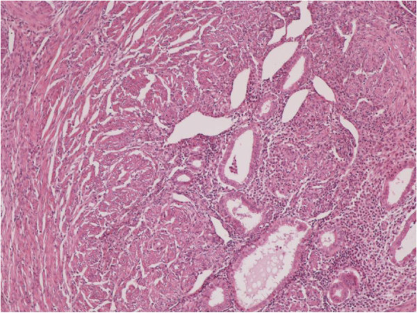
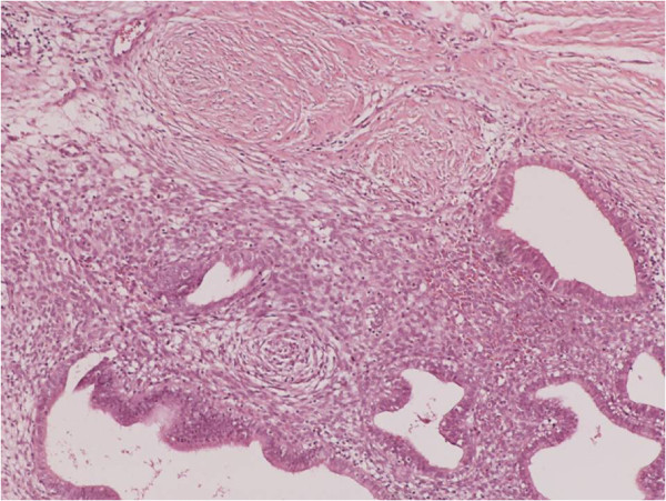
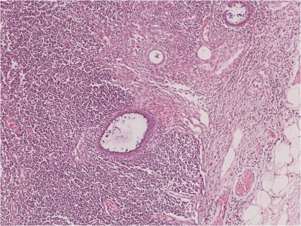
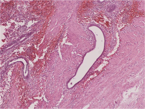
Endometriosis is a clinical entity characterized by the presence of normal endometrial mucosa abnormally implanted in locations other than the uterine cavity. Endometriosis can be either endopelvic or extrapelvic depending on the location of endometrial tissue implantation. Despite the rarity of extrapelvic endometriosis, several cases of endometriosis of the gastrointestinal tract, the urinary tract, the upper and lower respiratory system, the diaphragm, the pleura and the pericardium, as well as abdominal scars loci have been reported in the literature. There are several theories about the pathogenesis and the pathophysiology of endometriosis. Depending on the place of endometrial tissue implantation, endometriosis can be expressed with a wide variety of symptoms. The diagnosis of this entity is neither easy nor routine. Many diagnostic methods clinical and laboratory have been used, but none of them is the golden standard. The multipotent localization of endometriosis in combination with the wide range of its clinical expression should raise the clinical suspicion in every woman with periodic symptoms of extrapelvic organs. Finally, the therapeutic approach of this clinical entity is also correlated with the bulk of endometriosis and the locum that it is found. It varies from simple observation, to surgical treatment and treatment with medication as well as a combination of those. Virtual slides: The virtual slide(s) for this article can be found here: http://www.diagnosticpathology.diagnomx.eu/vs/1968087883113362.


Figures

Figure 1
Figure 1
Clusters of endometrial glands and stroma in fallopian tube wall with inflammation (H&E X100).
Figure 2
Figure 2
Nests of endometriosis into the rectus abdominis muscle (H&E X100).
Figure 3
Figure 3
Focal endometriosis in a lymph node (H&E X100).
Figure 4
Figure 4
Endometrial glands and stroma with hemorrhange and hemosiderin-laden macrophages into the muscularis propria of large bowel (H&E X100).
Figure 5
Figure 5
Nests of endometriosis into the muscularis propria of small bowel (H&E X40).
Figure 6
Figure 6
Endometrial glands and stroma into the dermis, close to cesarean section scar (H&E X100).
Figure 7
Figure 7
Nests of endometriosis in a fibrous backround, close to cesarean section scar (H&E X100).
Figure 8
Figure 8
MR images demonstrate foci of endometriomas at the sites of the section [T-2 w.i].
Figure 9
Figure 9
Endometrioma in the abdominal wall at point of the cesarian section [T-1w.i. and fat saturation technique].
Figure 10
Figure 10
Endometrioma after enhanced T-1 w.i. and fat saturation technique.
Figure 11
Figure 11
Hematoxylin and eosin (H&E). A: The alveolar spaces were filled with many red blood cells and phagocytic cells with hemosiderin (heavy arrowhead). The alveolar walls were infiltrated by plasma cells (triangulate arrowhead) and lymphocytes (light arrowhead); 200×. Immunohistochemical staining, B: Gland epithelium in an alveolus, CK7+ (heavy arrowhead). Infiltrating plasma cells and lymphocytes in the alveolar wall, CK7- (light arrowhead), H&E 200×, C: Phagocytic cells in the alveolar space, CD68+ (heavy arrowhead). Plasma cells and lymphocytes in the alveolar walls, CD68- (light arrowhead), H&E 200×. D: Atypical tubular-gland structures of decidual lesions were detected in the alveolar space (light arrowhead). Structure of alveolar wall (heavy arrowhead); 100×.

References

    1. Cassina PC, Hauser M, Kacl G, Imthurn B, Schroder S, Weder W. Catamenial hemoptysis. Diagnosis with MRI. Chest. 1997;8:1447–1450. doi: 10.1378/chest.111.5.1447. - DOI - PubMed
    1. Dragoumis K, Mikos T, Zafrakas M, Assimakopoulos E, Stamatopoulos P, Bontis J. Endometriotic uterocutaneous fistula after cesarean section. A case report. Gynecol Obstet Invest. 2004;8:90–92. doi: 10.1159/000075384. - DOI - PubMed
    1. Huanwen W, Hui Z, Xiaowei X, Zhaohui L. Extrauterine adenomyoma of the liver with a focally cellular smooth muscle component occurring in a patient with a history of myomectomy: case report and review of the literature. Diagn Pathol. 2013;8:131. doi: 10.1186/1746-1596-8-131. - DOI - PMC - PubMed
    1. Yu JH, Lin XY, Wang L, Liu Y, Fan CF, Zhang Y, Wang EH. Endobronchial endometriosis presenting as central-type lung cancer: a case report. Diagn Pathol. 2013;8:53. doi: 10.1186/1746-1596-8-53. - DOI - PMC - PubMed
    1. Uehara K, Yasuda M, Ichimura T, Yamaguchi H, Nagata K, Kayano H, Sasaki A, Murata S, Shimizu M. Peritoneal keratin granuloma associated with endometrioid adenocarcinoma of the uterine corpus. Diagn Pathol. 2011;8:104. doi: 10.1186/1746-1596-6-104. - DOI - PMC - PubMed