. 2014 Feb 15;458(1):41-56.
doi: 10.1042/BJ20131324.

Investigation of LKB1 Ser431 phosphorylation and Cys433 farnesylation using mouse knockin analysis reveals an unexpected role of prenylation in regulating AMPK activity

Affiliations

Investigation of LKB1 Ser431 phosphorylation and Cys433 farnesylation using mouse knockin analysis reveals an unexpected role of prenylation in regulating AMPK activity

Vanessa P Houde et al. Biochem J. .

Abstract

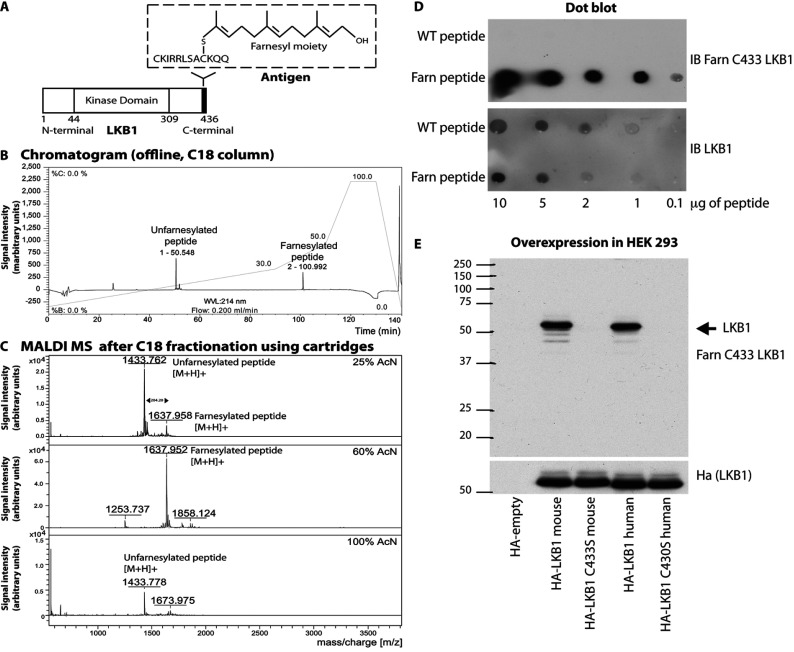
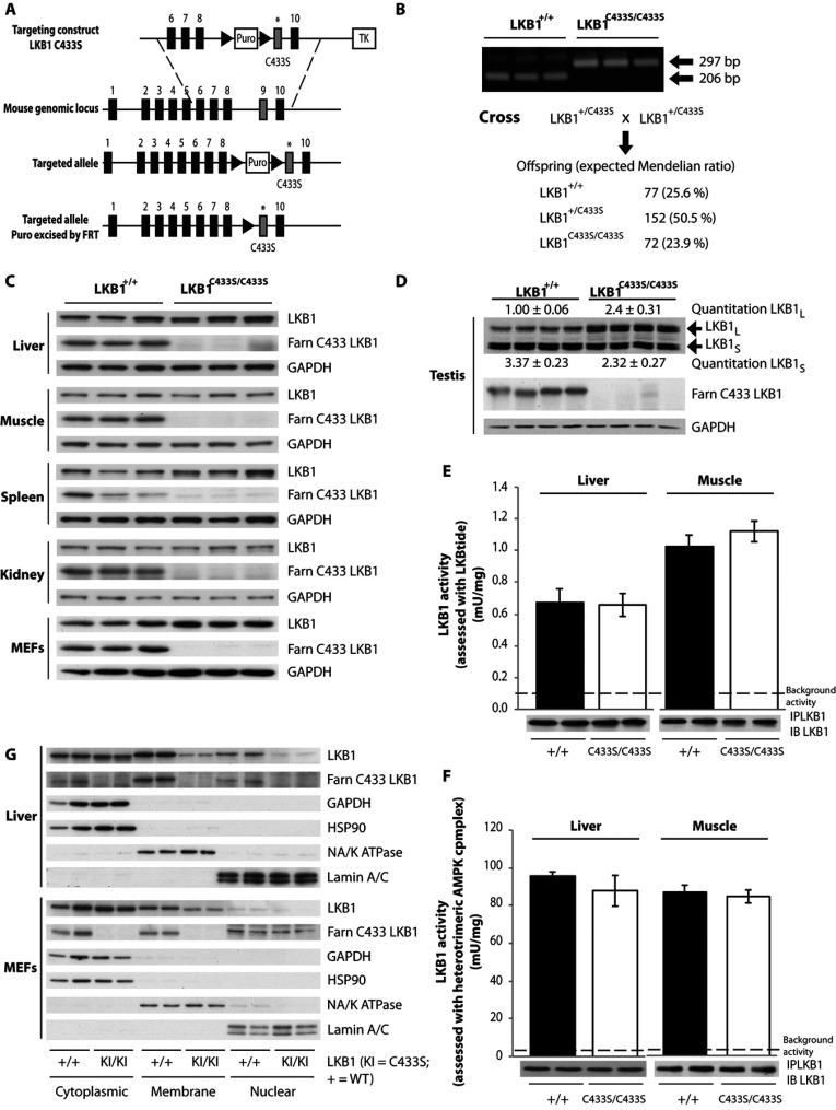
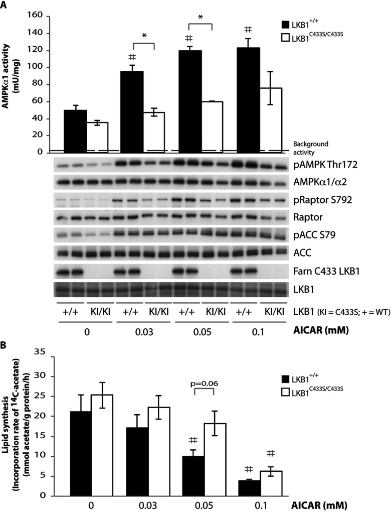
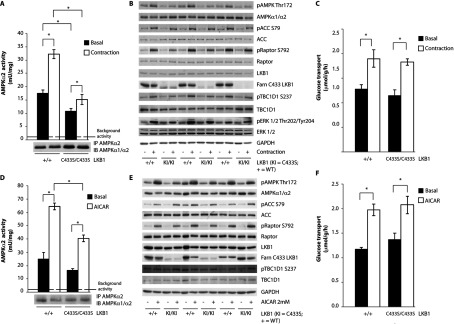
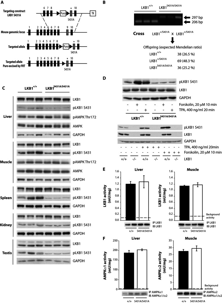
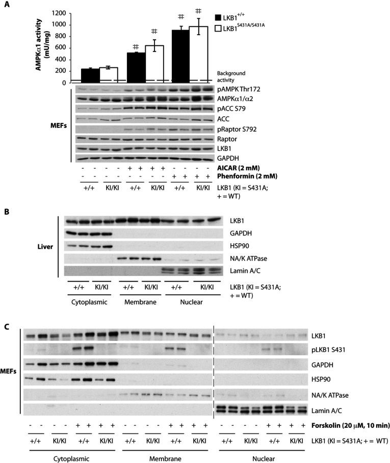
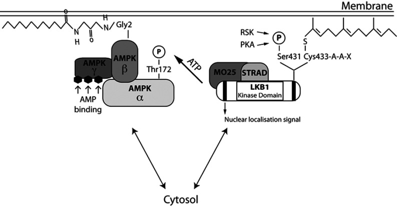
The LKB1 tumour suppressor protein kinase functions to activate two isoforms of AMPK (AMP-activated protein kinase) and 12 members of the AMPK-related family of protein kinases. The highly conserved C-terminal residues of LKB1 are phosphorylated (Ser431) by PKA (cAMP-dependent protein kinase) and RSK (ribosomal S6 kinase) and farnesylated (Cys433) within a CAAX motif. To better define the role that these post-translational modifications play, we created homozygous LKB1S431A/S431A and LKB1C433S/C433S knockin mice. These animals were viable, fertile and displayed no overt phenotypes. Employing a farnesylation-specific monoclonal antibody that we generated, we established by immunoprecipitation that the vast majority, if not all, of the endogenous LKB1 is prenylated. Levels of LKB1 localized at the membrane of the liver of LKB1C433S/C433S mice and their fibroblasts were reduced substantially compared with the wild-type mice, confirming that farnesylation plays a role in mediating membrane association. Although AMPK was activated normally in the LKB1S431A/S431A animals, we unexpectedly observed in all of the examined tissues and cells taken from LKB1C433S/C433S mice that the basal, as well as that induced by the AMP-mimetic AICAR (5-amino-4-imidazolecarboxamide riboside), AMPK activation, phenformin and muscle contraction were significantly blunted. This resulted in a reduced ability of AICAR to inhibit lipid synthesis in primary hepatocytes isolated from LKB1C433S/C433S mice. The activity of several of the AMPK-related kinases analysed [BRSK1 (BR serine/threonine kinase 1), BRSK2, NUAK1 (NUAK family, SNF1-like kinase 1), SIK3 (salt-inducible kinase 3) and MARK4 (MAP/microtubule affinity-regulating kinase 4)] was not affected in tissues derived from LKB1S431A/S431A or LKB1C433S/C433S mice. Our observations reveal for the first time that farnesylation of LKB1 is required for the activation of AMPK. Previous reports have indicated that a pool of AMPK is localized at the plasma membrane as a result of myristoylation of its regulatory AMPKβ subunit. This raises the possibility that LKB1 farnesylation and myristoylation of AMPKβ might promote the interaction and co-localization of these enzymes on a two-dimensional membrane surface and thereby promote efficient activation of AMPK.


Figures

Figure 1
Figure 1. Generation of a monoclonal antibody recognizing farnesylated LKB1
(A) Diagram illustrating the structure of the mouse LKB1 protein as well as the peptide used for the antibody production (antigen) with the farnesyl moiety attached to the C-terminal cysteine (Cys433). (B) HPLC chromatogram and (C) MALDI–TOF-MS of the peptide containing Cys433 before and after the farnesylation reaction. (D) Dot-blot analysis demonstrating that the anti-LKB1 farnesylated at Cys433 antibody does not recognize the C-terminal cysteine on the wild-type (WT) peptide (non-farnesylated peptide). Farn peptide, farnesylated peptide; IB, immunoblot. (E) HEK-293 cells were transfected with the indicated mouse and human HA–LKB1 mutants. The samples were immunoblotted with the anti-LKB1 farnesylated at Cys433 antibody to ensure that the antibody does not recognize the cysteine mutant proteins. Molecular mass is given on the left-hand side in kDa.
Figure 2
Figure 2. Characterization of the farnesylation-deficient LKB1C433S/C433S mouse
(A) The knockin construct, the endogenous LKB1 allele containing exons 1–10 and the target allele with the puromycin cassette (Puro) removed by Flp (flippase) recombinase are depicted. The black/grey rectangles represent exons and the black triangles represent FRT (flippase-recognition target) sites. TK, thymidine kinase. (B) To genotype mice, genomic DNA was PCR-amplified. Sizes are indicated in bp. The breeding strategy employed to generate LKB1C433S/C433S mice is shown with the number and percentage of each genotype obtained indicated. (C) Tissues lysates from wild-type LKB1+/+ and LKB1C433S/C433S mice and MEFs were subjected to immunoblotting using anti-LKB1 and anti-LKB1 farnesylation-specific (LKB1 Farn C433) antibodies. (D) Testis lysates from LKB1+/+ and LKB1C433S/C433S mice were immunoblotted with an anti-LKB1 antibody to detect the LKB1long (LKB1L) and LKB1short (LKB1s) isoforms. Band intensities were quantified using Li-Cor. Results are means±S.E.M. for four mice per genotype. (E) LKB1 was immunoprecipitated (IP) from the liver and muscle (EDL) from wild-type LKB1+/+ (+/+) and LKB1C433S/C433S (C433S/C433S) mice and the in vitro kinase activity towards the LKBtide peptide was measured. Immunoprecipitates were also immunoblotted (IB). Assays were performed in duplicate from tissues taken from six mice per genotype and the results are means±S.E.M. The broken line represents the background activity as measured with pre-immune IgG. (F) As (E), except that the immunoprecipitated LKB1 protein was used to activate a recombinant heterotrimeric AMPK complex (α1β2γ1) and then AMPK kinase activity towards the AMARA peptide was measured. Assays were performed in duplicate from tissues taken from six mice per genotype and the results are means±S.E.M. (G) Liver and MEFs from wild-type (WT) LKB1+/+ (+/+) and LKB1C433S/C433S (KI/KI) mice were submitted to subcellular fractionation. Cytoplasmic, membrane and nuclear fractions were immunoblotted with the indicated antibodies. Blots for two animals out of four per genotype are shown.
Figure 3
Figure 3. Impaired activity of AMPK and lipid synthesis in primary hepatocytes derived from LKB1C433S/C433S mice
Primary hepatocytes were isolated from wild-type LKB1+/+ (+/+) and LKB1C433S/C433S (KI/KI) mice and treated with increasing concentrations of AICAR for 1 h. (A) Upper panel: AMPKα1 was immunoprecipitated from primary hepatocytes extracts and the in vitro kinase activity towards the AMARA peptide was measured. Assays were performed in duplicate for each condition and results are means±S.E.M. for three independent experiments (n=3). For the non-stimulated conditions the P value of the data for LKB1+/+ compared with LKB1C433S/C433S mice was 0.1 and therefore judged not to be significant. The broken line represents the background activity as measured with pre-immune IgG. Lower panel: primary hepatocyte extracts were immunoblotted with the indicated antibodies. Representative immunoblots for three independent experiments (n=3) are shown. LKB1 Farn C433, anti-LKB1 farnesylation-specific antibody. (B) Lipogenesis following AICAR treatment in primary hepatocytes was assessed by using [14C]acetate incorporation. Assays were performed in triplicate for each condition and results are means±S.E.M. for four independent experiments (n=4). *P<0.05 LKB1+/+ compared with LKB1C433S/C433S mice within each condition and #P<0.05 treatment compared with non-treatment condition. Statistical analysis was performed using one-way ANOVA and Tukey's post-hoc test.
Figure 4
Figure 4. Impaired activity of AMPK in muscles taken from LKB1C433S/C433S mice
(A) One leg from anaesthetized wild-type LKB1+/+ (+/+) and LKB1C433S/C433S (C433S/C433S) mice was subjected to in situ hindlimb contraction (Contraction) via sciatic nerve stimulation for 5 min and the other leg served as a non-contracted control (Basal). AMPKα2 was immunoprecipitated (IP) from tibialis anterior lysates and the in vitro kinase activity towards the AMARA peptide was measured. The immunoprecipitates were also immunoblotted (IB). Assays were performed in duplicate from lysates derived from five mice per genotype and results are means±S.E.M. The broken line represents the background activity as measured with pre-immune IgG. (B) Tibialis anterior muscle lysates were submitted to immunoblotting with the indicated antibodies. A total of three animals out of five per genotype are shown (n=5). (C) Following contraction, EDL muscles were isolated and glucose transport was measured. Results are means±S.E.M. (n=4–7 per group). (D) Isolated EDL muscle taken from wild-type LKB1+/+ (+/+) and LKB1C433S/C433S (C433S/C433S) mice were incubated in the presence or absence of 2 mM AICAR for 50 min. AMPKα2 was immunoprecipitated from EDL lysates and the in vitro kinase activity towards the AMARA peptide was measured. The immunoprecipitates were also immunoblotted. Assays were performed in duplicate from lysates derived from four to five mice per genotype and results are means±S.E.M. The broken line represents the background activity as measured with pre-immune IgG. (E) EDL muscle lysates were submitted to immunoblotting with the indicated antibodies. A total of three animals out of five per genotype are shown (n=5). (F) Glucose transport in isolated EDL muscle was measured. Results are means±S.E.M. (n=4–7 per group). *P<0.05 basal compared with contraction or AICAR stimulation within each genotype. Statistical analysis was performed using one-way ANOVA and Tukey's post-hoc test.
Figure 5
Figure 5. Characterization of the LKB1S431A/S431A mouse
(A) The knockin construct, the endogenous LKB1 allele containing exons 1–10 and the target allele with the puromycin cassette (Puro) removed by Flp recombinase are shown. The black/grey rectangles represent exons and the black triangles represent FRT (flippase-recognition target) sites. TK, thymidine kinase. (B) To genotype mice, genomic DNA was PCR-amplified. Sizes are indicated in bp. The breeding strategy employed to generate LKB1S431A/S431A mice is shown with the number and percentage of each genotype obtained indicated. (C) Tissues lysates from wild type LKB1+/+ and LKB1S431A/S431A mice were subjected to immunoblotting using anti-LKB1 and anti-phospho-LKB1 Ser431 antibodies. (D) MEFs derived from wild-type LKB1+/+ and LKB1S431A/S431A mice (upper panel) or wild type LKB1+/+ (+/+) and knockout LKB1−/− (−/−) mice (lower panel) were stimulated with forskolin (20 μM for 10 min) or TPA (400 ng/ml for 20 min) and immunoblotted with the indicated antibodies. (E) LKB1 was immunoprecipitated (IP) from the liver and muscle (EDL) from wild-type LKB1+/+ (+/+) and LKB1S431A/S431A (S431A/S431A) mice and the in vitro kinase activity towards the LKBtide peptide was measured. Immunoprecipitates were also immunoblotted (IB). Assays were performed in duplicate from tissues derived from four mice per genotype and results are means±S.E.M. The broken line represents the background activity as measured with pre-immune IgG. (F) AMPKα1 (liver) and AMPKα2 (muscle; EDL) were immunoprecipitated from wild-type LKB1+/+ (+/+) and LKB1S431A/S431A (S431A/S431A) mice and the in vitro kinase activity towards the AMARA peptide was measured. Immunoprecipitates were also immunoblotted. Assays were performed in duplicate from tissues derived from four mice per genotype and results are means±S.E.M. The broken line represents the background activity as measured with pre-immune IgG.
Figure 6
Figure 6. Normal activity of AMPK in MEFs derived from wild-type LKB1+/+ (+/+) and LKB1S431A/S431A (KI/KI) mice
(A) MEFs were treated with 2 mM AICAR or phenformin for 1 h. AMPKα1 was immunoprecipitated from MEF extracts and the in vitro kinase activity towards the AMARA peptide was measured. Assays were performed in duplicate for each condition and results are means±S.E.M. The broken line represents the background activity as measured with pre-immune IgG. MEF extracts were immunoblotted with the indicated antibodies. No significant basal phosphorylation was observed for the Ser431 site in the LKB1S431A/S431A MEFs (results not shown). #P<0.05 treated compared with non-treated cells. Statistical analysis was performed using one-way ANOVA and Tukey's post-hoc test. There was a normal localization of LKB1 in the liver or MEFs derived from wild-type LKB1+/+ and LKB1S431A/S431A mice. Livers (B) and MEFs (C) from wild-type LKB1+/+ (+/+) and LKB1S431A/S431A (KI/KI) mice were submitted to subcellular fractionation. Cytoplasmic, membrane and nuclear fractions were immunoblotted with the indicated antibodies. No significant basal phosphorylation was observed for the Ser431 site in the LKB1S431A/S431A liver fractions (results not shown). The broken line indicates that the samples were processed in parallel on different gels.
Figure 7
Figure 7. Schematic representation of the potential mechanism by which LKB1 farnesylation and myristoylation of AMPKβ might operate to promote the interaction of LKB1 and AMPK by localizing these enzymes on a two-dimensional membrane surface
The LKB1–STRAD–MO25 complex is anchored to the membrane through an LKB1 farnesylation motif. AMPKβ myristoylation promotes the kinase association to the membrane. In the presence of an increased ratio of AMP/ATP or other metabolic signals that deplete the intracellular ATP levels, the co-localization of these two kinases at the membrane allows LKB1 to phosphorylate AMPK on its T-loop residue (Thr172) and thereby fully activates AMPK in response to a metabolic signal.

References

    1. Hemminki A., Markie D., Tomlinson I., Avizienyte E., Roth S., Loukola A., Bignell G., Warren W., Aminoff M., Hoglund P., et al. A serine/threonine kinase gene defective in Peutz–Jeghers syndrome. Nature. 1998;391:184–187. - PubMed
    1. Sanchez-Cespedes M. A role for LKB1 gene in human cancer beyond the Peutz–Jeghers syndrome. Oncogene. 2007;26:7825–7832. - PubMed
    1. Ji H., Ramsey M. R., Hayes D. N., Fan C., McNamara K., Kozlowski P., Torrice C., Wu M. C., Shimamura T., Perera S. A., et al. LKB1 modulates lung cancer differentiation and metastasis. Nature. 2007;448:807–810. - PubMed
    1. Sanchez-Cespedes M., Parrella P., Esteller M., Nomoto S., Trink B., Engles J. M., Westra W. H., Herman J. G., Sidransky D. Inactivation of LKB1/STK11 is a common event in adenocarcinomas of the lung. Cancer Res. 2002;62:3659–3662. - PubMed
    1. Baas A. F., Boudeau J., Sapkota G. P., Smit L., Medema R., Morrice N. A., Alessi D. R., Clevers H. C. Activation of the tumour suppressor kinase LKB1 by the STE20-like pseudokinase STRAD. EMBO J. 2003;22:3062–3072. - PMC - PubMed

Publication types