. 2013 Dec 2;10(1):19.
doi: 10.1186/1559-0275-10-19.

Chitotriosidase - a putative biomarker for sporadic amyotrophic lateral sclerosis

Affiliations

Chitotriosidase - a putative biomarker for sporadic amyotrophic lateral sclerosis

Anu Mary Varghese et al. Clin Proteomics. .

Abstract

Background: Potential biomarkers to aid diagnosis and therapy need to be identified for Amyotrophic Lateral Sclerosis, a progressive motor neuronal degenerative disorder. The present study was designed to identify the factor(s) which are differentially expressed in the cerebrospinal fluid (CSF) of patients with sporadic amyotrophic lateral sclerosis (SALS; ALS-CSF), and could be associated with the pathogenesis of this disease.

Results: Quantitative mass spectrometry of ALS-CSF and control-CSF (from orthopaedic surgical patients undergoing spinal anaesthesia) samples showed upregulation of 31 proteins in the ALS-CSF, amongst which a ten-fold increase in the levels of chitotriosidase-1 (CHIT-1) was seen compared to the controls. A seventeen-fold increase in the CHIT-1 levels was detected by ELISA, while a ten-fold elevated enzyme activity was also observed. Both these results confirmed the finding of LC-MS/MS. CHIT-1 was found to be expressed by the Iba-1 immunopositive microglia.

Conclusion: Elevated CHIT-1 levels in the ALS-CSF suggest a definitive role for the enzyme in the disease pathogenesis. Its synthesis and release from microglia into the CSF may be an aligned event of neurodegeneration. Thus, high levels of CHIT-1 signify enhanced microglial activity which may exacerbate the process of neurodegeneration. In view of the multifold increase observed in ALS-CSF, it can serve as a potential CSF biomarker for the diagnosis of SALS.


Figures
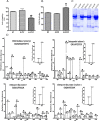
Figure 1
Figure 1
Mass spectrometric analysis of ALS-CSF samples: Toxicity assays (A & B) were performed. Histogram of MTT assay showing 40% reduction in the viability of NSC-34 cells upon exposure to 10%(v/v) ALS-CSF compared to the cells exposed to normal-CSF (###p < 0.001 vs. N-CSF) and 30% compared to normal controls (***p < 0.001 vs. NC; A). ALS-CSF caused significant increase in LDH activity when compared to control groups (***p < 0.001 vs. NC and $$$p < 0.001 vs. N-CSF; B). Gel image representing depletion of abundant proteins prior to mass spectrometric analysis (C). Representative MS/MS spectra of the peptides of CHIT1, Osteopontin, CHI3L1 and CHI3L2 (D). Tests of significance was Student’s t test, One way Anova followed by Tukey’s post hoc analysis.
Figure 2
Figure 2
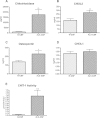
Validation of upregulated proteins by ELISA and CHIT-1 activity assay. Chitotriosidase was found to be 17 fold upregulated (**p < 0.01 vs. N-CSF; A) and chitinase 3- like-2 (CHI3L2) showed ~2 fold increase (**p < 0.01 vs. N-CSF; B) in ALS-CSF. Osteopontin showed ~ 1.9 fold upregulation (*p < 0.05 vs. N-CSF; C) whereas, upregulation in Chitinase 3- like-1 (CHI3L1) levels was statistically non- significant (D). Histogram showing CHIT-1 enzymatic activity in CSF samples. The activity was found to be 10 fold higher in ALS-CSF (**p < 0.001 vs. N-CSF; E).
Figure 3
Figure 3
Expression of CHIT-1 in microglial cultures. Immunoflourescence photomicrographs of pure microglial cultures labeled with CHIT-1 (green; A, D) and Iba-1, a marker for microglia (red; B, E). Note the increased expression of CHIT-1 in the cultures exposed to ALS-CSF (D) as compared to the normal control (A). The merged images (C and F) depict the co-labeling of CHIT-1 with Iba-1 in normal control (A) and ALS group (D) respectively.
Figure 4
Figure 4
A schematic representation of the procedural steps for mass spectrometric analysis. Pooled CSF samples were subjected to mass spectrometry after sequential treatment procedures like, depletion of abundant proteins, tryptic digestion, iTRAQ labelling and SCX. Selected molecules were validated using ELISA.

References

    1. Wagner KR. The need for biomarkers in amyotrophic lateral sclerosis drug development. Neurology. 2009;72:11–12. doi: 10.1212/01.wnl.0000338538.18938.9d. - DOI - PubMed
    1. Shaw C. What have cellular models taught us about ALS? Amyotroph Lateral Scler Other Motor Neuron Disord. 2002;3:55–56. - PubMed
    1. Anneser JM, Chahli C, Borasio GD. Protective effect of metabotropic glutamate receptor inhibition on amyotrophic lateral sclerosis-cerebrospinal fluid toxicity in vitro. Neuroscience. 2006;141:1879–1886. doi: 10.1016/j.neuroscience.2006.05.044. - DOI - PubMed
    1. Shahani N, Nalini A, Gourie-Devi M, Raju TR. Reactive astrogliosis in neonatal rat spinal cord after exposure to cerebrospinal fluid from patients with amyotrophic lateral sclerosis. Exp Neurol. 1998;149:295–298. doi: 10.1006/exnr.1997.6651. - DOI - PubMed
    1. Shahani N, Gourie-Devi M, Nalini A, Raju TR. Cyclophosphamide attenuates the degenerative changes induced by CSF from patients with amyotrophic lateral sclerosis in the neonatal rat spinal cord. J Neurol Sci. 2001;185:109–118. doi: 10.1016/S0022-510X(01)00479-8. - DOI - PubMed