

Review
doi: 10.1101/cshperspect.a008987.
Molecular mechanisms of SH2- and PTB-domain-containing proteins in receptor tyrosine kinase signaling
Affiliations
- PMID: 24296166
- PMCID: PMC3839611
- DOI: 10.1101/cshperspect.a008987
Item in Clipboard
Review
Molecular mechanisms of SH2- and PTB-domain-containing proteins in receptor tyrosine kinase signaling
Cold Spring Harb Perspect Biol.
.
Abstract
Intracellular signaling is mediated by reversible posttranslational modifications (PTMs) that include phosphorylation, ubiquitination, and acetylation, among others. In response to extracellular stimuli such as growth factors, receptor tyrosine kinases (RTKs) typically dimerize and initiate signaling through phosphorylation of their cytoplasmic tails and downstream scaffolds. Signaling effectors are recruited to these phosphotyrosine (pTyr) sites primarily through Src homology 2 (SH2) domains and pTyr-binding (PTB) domains. This review describes how these conserved domains specifically recognize pTyr residues and play a major role in mediating precise downstream signaling events.
Figures
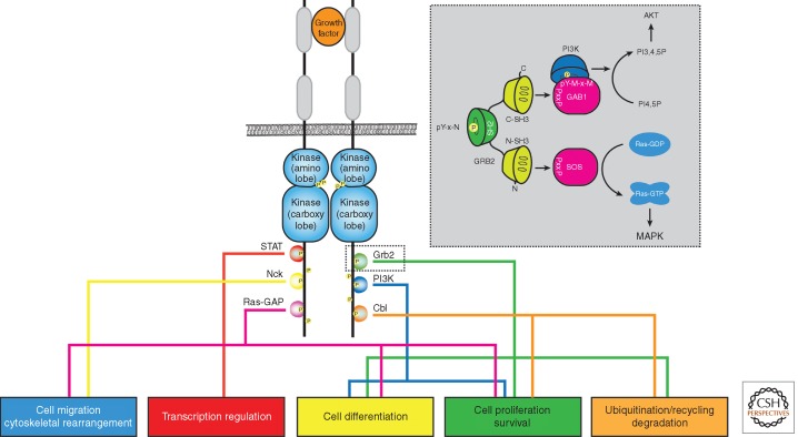
Receptor tyrosine kinases activate downstream pathways through recruitment of proteins containing pTyr-binding domains. Receptor tyrosine kinases are activated on growth factor binding to the extracellular domain of the receptor, leading to receptor dimerization and tyrosine phosphorylation (yellow circles labeled with a P) of their cytoplasmic tails, which act as docking sites for recruitment of PTB and SH2 domains. Various RTKs can mediate a diverse set of cellular processes (colored boxes) determined by the recruitment of specific SH2- and PTB-domain-containing proteins. The gray box displays how the adaptor Grb2 is recruited to an RTK through recognition of the pY-x-N (pY = pTyr, x = any natural amino acid) and activates cell growth and survival pathways such as MAPK and AKT, respectively, through complex formation via its SH3 domains.
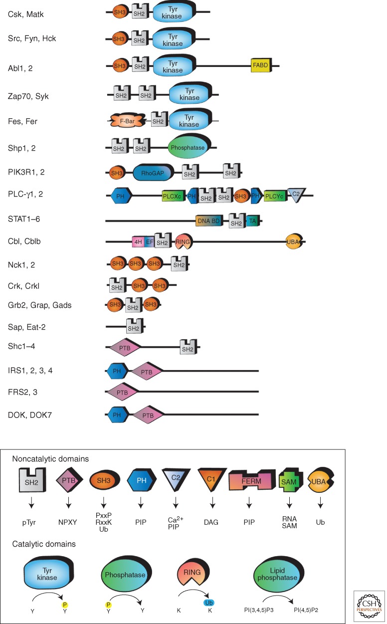
SH2- and PTB-containing proteins are diverse in nature. The modular domain organization of SH2- and PTB-domain proteins displays a diverse set of noncatalytic and catalytic domains for mediating protein–protein interactions and enzyme catalysis, respectively. See the box legend for description of the binding partners and function of these domains. More information on the individual domains portrayed can be found at www.pawsonlab.mshri.on.ca and www.smart.embl-heidelberg.de .
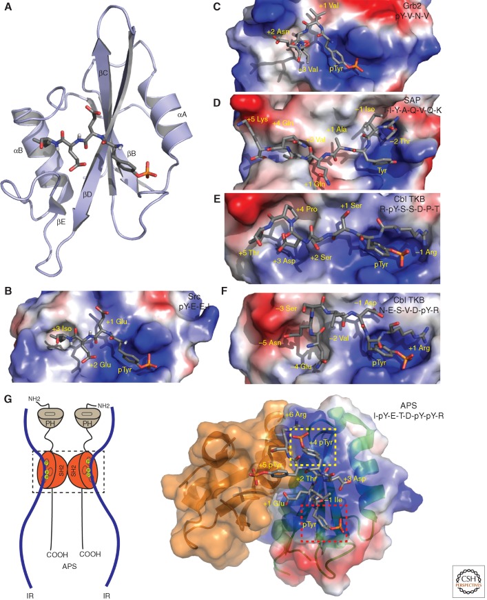
Src homology 2 domains recognize specific phosphotyrosine motifs. (A) Ribbon structure of the Src SH2 domain (light blue) bound to a pTyr-Glu-Glu-Ile peptide (PDB: 1SPS) (Waksman et al. 1993). The amino-terminal pTyr of the peptide (gray) occupies the pTyr-binding pocket. The peptide runs over the central β sheet of the SH2 domain, the +1 and +2 glutamates contact the surface of the domain, and the side chain of the +3 Ile (to the left) fits in a hydrophobic pocket. (B) The electrostatic surface of the SH2 domain reveals the positive charged pTyr-binding pocket (blue is positive, red is negative) and the ligand-binding pocket. (C) Grb2 SH2 domain in complex with pYVNV (red) (PDB: 1BMB). (D) The SAP SH2 domain can recognize nonphoshorylated SLAM peptide and residues amino terminal to the tyrosine, such as -2 Thr (PDB: 1M27). (E) pTyr-binding pocket of the Cbl SH2 is bound in the canonical fashion with EGFR peptide pYSSDP (gray) with the carboxyl terminus extended across the SH2 surface (PDB: 3BUO). (F) Cbl TKB in complex with the MET peptide is oriented in the reverse direction with the amino acids amino terminal to the pTyr extended across the SH2 domain (PDB: 3BUX). (G) Left panel: A graphical representation of the dimerized APS molecules bound to the insulin receptor (IR). Right panel: The dimerized SH2 domain of APS bound to the activation loop peptide of the IR with the primary pTyr pocket (red box) and the second pocket (yellow box) indicated.
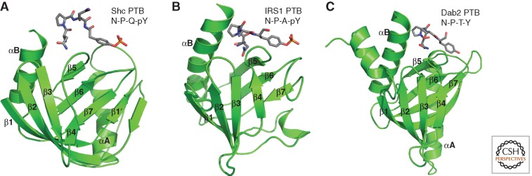
Structure of phosphotyrosine-binding domains. The ribbon structures shown in green of the PTB domains of Shc (A) (PDB: 1SHC), IRS-1 (B) (PDB: 1IRS), and Dab2 (C) (PTB: 1ME7) bound to their respective N-P-x-(p)Y ligand (gray). x Denotes any natural amino acid; pY represents phosphotyrosine. The mode of ligand binding to the PTB domains shown is similar.
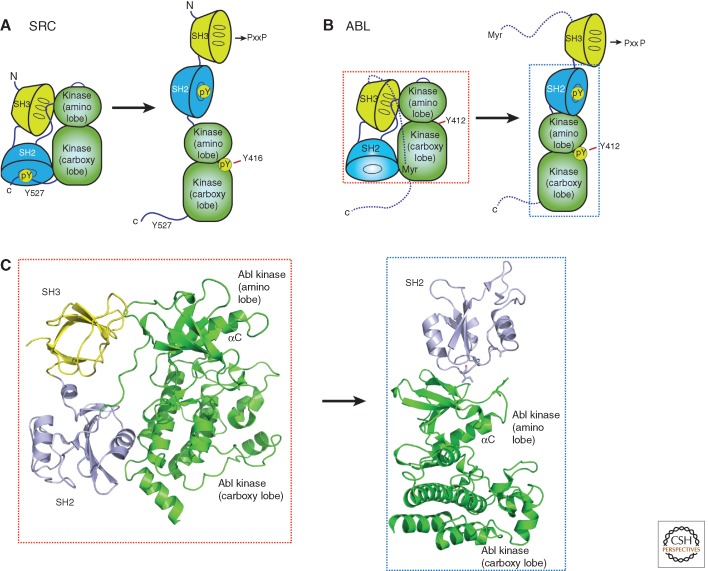
SH2 domains regulate tyrosine kinase activation. (A) In the autoinhibited state of Src, the SH2 domain recognizes an intramolecular pTyr site (Y572) and stabilizes the inactive kinase. Additional contacts with the SH3 and the SH2-kinase linker promote stabilization of the inactive conformation of Src. Activation of Src, initiated by dephosphorylation of Y527, frees the SH3 and SH2 to recognize other short linear motifs and further promote kinase activity. (B) The Abl tyrosine kinase remains in an inactive conformation analogous to Src but lacks the intramolecular phosphorylation site. Instead, an intramolecular amino-terminal myristolation stabilizes the inactive conformation of Abl. (C) For Abl kinase to be active, it requires the SH2 domain to stabilize the amino lobe of the kinase domain, thereby allowing it to couple pTyr ligand binding and substrate recognition (PDB:1OPL).
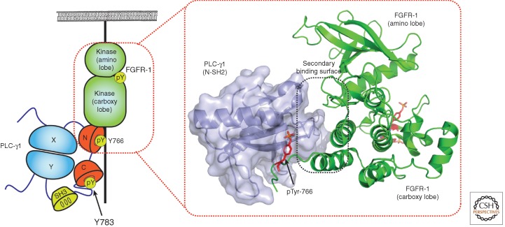
Activation of PLC-γ. Phospholipase C isoform γ (PLC-γ1) is a tandem SH2 domain-containing protein (see Fig. 2 for a complete view of the domain organization of PLC-γ1) that is recruited to activated FGFR-1. The amino-terminal SH2 domain of PLC-γ1 mediates primary pTyr binding to Y766 on FGFR-1 but also mediates secondary contacts through the B,C loop and D,E loops of the SH2 domain and the αE, I helix of the carboxy lobe of the kinase domain (PDB: 3GQI). In the activated state of PLC-γ1, the carboxy-terminal SH2 recognizes an intramolecular tyrosine phosphorylation site Y783 bringing together the PLC-γ-x (X) and PLC-γ-y (Y) lobes, activating the phospholipase enzyme.
References
-
- Anderson D, Koch CA, Grey L, Ellis C, Moran MF, Pawson T 1990. Binding of SH2 domains of phospholipase Cγ1, GAP, and Src to activated growth factor receptors. Science 250: 979–982 - PubMed
-
- Araki T, Nawa H, Neel BG 2003. Tyrosyl phosphorylation of Shp2 is required for normal ERK activation in response to some, but not all, growth factors. J Biol Chem 278: 41677–41684 - PubMed
Publication types
MeSH terms
Substances
LinkOut - more resources
Full Text Sources
Other Literature Sources
Miscellaneous