

Review
doi: 10.1101/cshperspect.a009233.
Central role of RET in thyroid cancer
Affiliations
- PMID: 24296167
- PMCID: PMC3839608
- DOI: 10.1101/cshperspect.a009233
Item in Clipboard
Review
Central role of RET in thyroid cancer
Cold Spring Harb Perspect Biol.
.
Abstract
RET (rearranged during transfection) is a receptor tyrosine kinase involved in the development of neural crest derived cell lineages, kidney, and male germ cells. Different human cancers, including papillary and medullary thyroid carcinomas, lung adenocarcinomas, and myeloproliferative disorders display gain-of-function mutations in RET. Accordingly, RET protein has become a promising molecular target for cancer treatment.
Figures
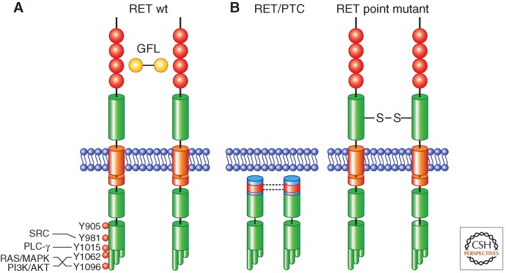
Illustration of the mechanisms of activation of wild-type (wt) RET and RET-derived oncoproteins. (A) Wild-type RET activation is mediated by ligand (GFL)-induced dimerization; ligand binding to RET is not direct and mediated by GFR-α coreceptors (not shown); major RET autophosphorylation sites and downstream signaling pathways are indicated. RET extracellular cadherin-like domains are represented in red. The split intracellular RET tyrosine kinase domain, as well as the three alternative carboxy-terminal RET tails, are also depicted. (B) RET/PTC activation is mediated by coiled-coil-induced dimerization (left); activation of RET cysteine mutants associated with MEN2A or FMTC is mediated by disulfide bonds-mediated dimerization (right).
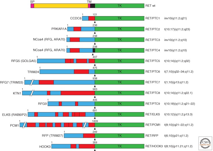
Schematic representation of RET/PTC oncoproteins. On the top, wild-type RET protein is illustrated. For each RET/PTC rearrangement, the name of the fusion partner is indicated on the left and the corresponding chromosomal alteration is indicated on the right. The fusion points are indicated by arrowheads. The length in amino acids of the partner protein portion is also indicated. Boxes in red indicate dimerization (coiled-coil) domains. SP, Signal peptide; TM, transmembrane domain; TK, tyrosine kinase domain; CCDC6, coiled-coil domain-containing protein 6; PRKAR1A, protein kinase, cAMP-dependent, regulatory, type I, alpha; NCOA4 (RFG, ARA70), nuclear coactivator 4 (RET-fused gene, androgen receptor-associated protein 70); RFG5 (GOLGA5), RET-fused gene 5 (Golgin A5); TRIM24, tripartite motif-containing 24; RFG7 (TRIM33), RET-fused gene 7 (tripartite motif-containing 33); KTN1, kinectin 1; RFG9, RET-fused gene 9; ELKS (RAB6IP2), glutamate, leucine, lysine, serine-rich sequence (RAB6-interacting protein 2); PCM1, pericentriolar material 1; RFP (TRIM27), RET finger protein (tripartite motif-containing 27); HOOK3, hook homolog 3.
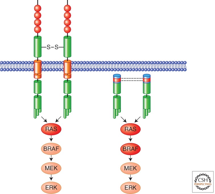
RAS-MAPK oncogenic signaling cascade activated by RET oncoproteins in MTC (left) and PTC (right). In red are indicated other mutational targets (RAS and BRAF) associated with MTC or PTC, respectively. In most of the cases, these mutations are alternative and not present simultaneously in a single tumor.
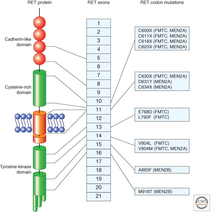
Representation of the most common germline missense mutations in RET gene found in MEN2 syndromes. The location of the mutation with respect to RET protein domains and RET exons is indicated. MEN2 phenotypes associated with the various mutations are indicated on the right.
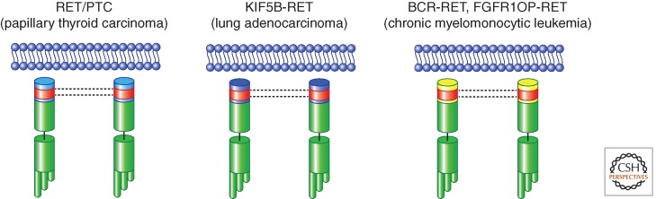
Schematic representation of RET gene rearrangements in human cancer. RET/PTC rearrangements in PTC are shown, together with KIF5B-RET (found in lung adenocarcinoma) and BCR-RET and FGFR1OP-RET (found in chronic myelomonocytic leukemia). All the RET fusion partners invariably code for protein–protein dimerization motifs (depicted as red boxes).
References
-
- Airaksinen MS, Saarma M 2002. The GDNF family: Signalling, biological functions and therapeutic value. Nat Rev Neurosci 3: 383–394 - PubMed
-
- Alberti L, Borrello MG, Ghizzoni S, Torriti F, Rizzetti MG, Pierotti MA 1998. Grb2 binding to the different isoforms of Ret tyrosine kinase. Oncogene 17: 1079–1087 - PubMed
-
- Ameziane-El-Hassani R, Boufraqech M, Lagente-Chevallier O, Weyemi U, Talbot M, Métivier D, Courtin F, Bidart JM, El Mzibri M, Schlumberger M, et al. 2010. Role of H2O2 in RET/PTC1 chromosomal rearrangement produced by ionizing radiation in human thyroid cells. Cancer Res 70: 4123–4132 - PubMed
-
- Anders J, Kjar S, Ibáñez CF 2001. Molecular modeling of the extracellular domain of the RET receptor tyrosine kinase reveals multiple cadherin-like domains and a calcium-binding site. J Biol Chem 276: 35808–35817 - PubMed
-
- Arighi E, Popsueva A, Degl’Innocenti D, Borrello MG, Carniti C, Perälä NM, Pierotti MA, Sariola H 2004. Biological effects of the dual phenotypic Janus mutation of ret cosegregating with both multiple endocrine neoplasia type 2 and Hirschsprung’s disease. Mol Endocrinol 18: 1004–1017 - PubMed
Publication types
MeSH terms
Substances
Supplementary concepts
LinkOut - more resources
Full Text Sources
Other Literature Sources
Medical