Review
. 2013 Dec;15(12):1387-97.
doi: 10.1038/ncb2888.

The roles of evolutionarily conserved functional modules in cilia-related trafficking

Affiliations
Review

The roles of evolutionarily conserved functional modules in cilia-related trafficking

Ching-Hwa Sung et al. Nat Cell Biol. 2013 Dec.

Abstract

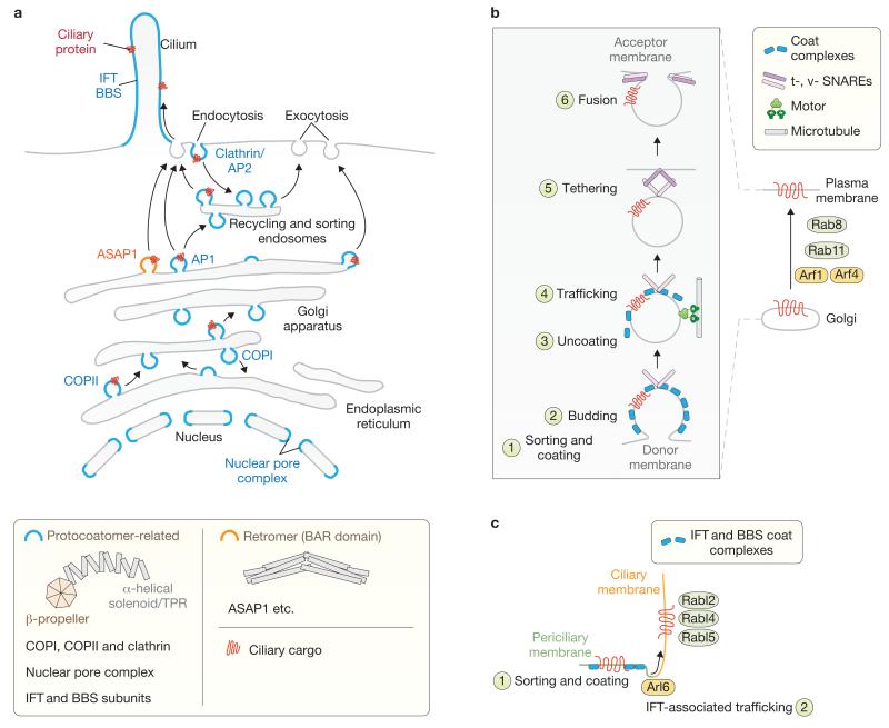
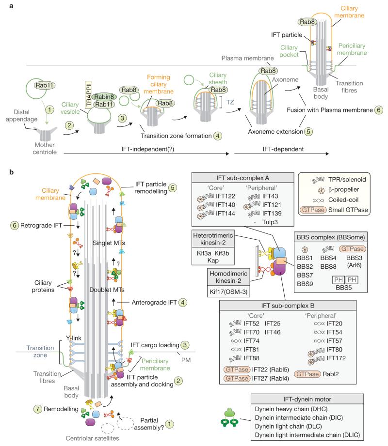
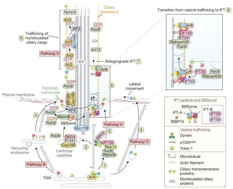
Cilia are present across most eukaryotic phyla and have diverse sensory and motility roles in animal physiology, cell signalling and development. Their biogenesis and maintenance depend on vesicular and intraciliary (intraflagellar) trafficking pathways that share conserved structural and functional modules. The functional units of the interconnected pathways, which include proteins involved in membrane coating as well as small GTPases and their accessory factors, were first experimentally associated with canonical vesicular trafficking. These components are, however, ancient, having been co-opted by the ancestral eukaryote to establish the ciliary organelle, and their study can inform us about ciliary biology in higher organisms.


Figures

Figure 1
Figure 1
Functional modules used in membrane trafficking and shaping already established in the last eukaryotic common ancestor (LECA). (a) Membrane-coating modules in the eukaryotic cell implicated in vesicular trafficking, intraflagellar transport (IFT) and nuclear pore complex (NPC) formation. Two major classes of membrane-coating or deforming systems are represented: the protocoatomer-related and the retromer (BAR-domain-containing) complexes. Notably, COPI, COPII and clathrin coats as well as NPC and IFT (BBS) subunits share an evolutionarily conserved β-propeller–solenoid/TPR structural architecture formed from one or two polypeptides. A few key vesicular and intraciliary trafficking components are noted. (b) General mechanisms of vesicular trafficking and regulation by small GTPases. Right: a host of small GTPases (for example, Rab8, Rab11, Arf1 and Arf4) mobilize membrane-associated cargo from the Golgi to the plasma membrane. Left: a more detailed schematic of membrane trafficking. The sorting of cargo and coating of vesicles (1) and budding (2) steps are shown before uncoating and motor-dependent trafficking events (3,4) that ultimately lead to the tethering of the vesicle via t- and v-SNAREs and fusion with the acceptor membrane (5,6). Not shown are various adaptors and tethers, and involvement of the exocyst complex in the final stage of trafficking. (c) Non-vesicular-based mechanism for the trafficking of a protein from the periciliary membrane into the ciliary compartment. Sorting and coating by BBS coat complexes (1) uses the small GTPase Arl6 (BBS3) and is coordinated with the IFT trafficking machinery to move proteins into the cilium (2), with the assistance of Rab-like (Rabl) small GTPases. Sorting and coating by IFT proteins probably occurs in parallel.
Figure 2
Figure 2
Ciliary-vesicle-dependent steps of ciliogenesis, and modular organization and mechanism of the IFT machinery. (a) A pathway for ciliogenesis involves a ciliary vesicle and the small GTPases Rab8 and Rab11. The mother centriole uses distal appendages (which mature into transition fibres) to interact with a Rab11-associated ciliary vesicle (1). Rabin8 and coat protein small GTPase tethering complexes (including TRAPPII) are recruited to the ciliary vesicle (2). Rab8 is then recruited and seems to mark a Rab11-to-Rab8 switch, although this event is not clearly defined spatiotemporally (3). A transition zone (TZ) emerges (4), from which axoneme extension occurs (5), either before or following fusion of the invaginated ciliary vesicle with the plasma membrane (6). Both the transition fibres on the basal body and mature TZ help to seal the ciliary compartment, which is identifiable by the presence of Rab8. (b) Model for IFT-mediated transport. A complete anterograde IFT particle (including kinesin motor(s), IFT sub-complexes A and B, and the BBSome) assembles near or at the basal body transition fibres in association with the periciliary membrane, from components trafficked to the base of the cilium and centriolar satellites (1,2). Ciliary cargo is loaded onto IFT particles (3) and transported to the tip of the cilium (4) using one or more anterograde motors. Heterotrimeric kinesin-2 (Kin-II) is normally required for this motility, although additional kinesins may be used, including Kif17 (OSM-3). Remodelling at the tip (5) prepares the IFT machinery for retrograde transport (6), and at the base completes the IFT cycle (7). How the dynein and kinesin machineries move to the tip and back, respectively, remains unclear (shown as question marks). Cilia possess a proximal axoneme composed of doublet microtubules (MTs), and often have a distal axoneme with singlet MTs. In many C. elegans cilia, Kin-II and OSM-3 operate coordinately along doublet MTs, whereas OSM-3 acts alone in the distal segment. On the right, the composition and organization of the IFT machinery are depicted together with their various structural and functional domains (including β-propeller and TPR/solenoid motifs, small GTPases, coiled-coils and pleckstrin homology (PH) phosphatidylinositol lipid-binding module).
Figure 3
Figure 3
Model depicting ciliary cargo trafficking pathways. Cilium-bound, membrane-associated proteins are initially sorted at the trans-Golgi network (TGN) before being delivered to the subapical (pericentriolar) region. There is evidence for at least four possible transport pathways. Pathway A involves direct targeting to the periciliary membrane. At the TGN, Arf4 regulates the budding of vesicles containing membrane-associated ciliary proteins, and Tctex-1 (Dynlt1) binds to a ciliary targeting sequence on the cytoplasmic domains of a cargo protein (for example, rhodopsin) to promote dynein motor coupling (1). Additional regulatory components, including Rab8, Rab11, Asap1 and FIP3 (not shown), and at least one IFT protein (IFT20), may also ride on the pericentriolarly directed transport vesicles (2). Centriolar satellites associated with the basal body (3) may act as a way station for ciliary trafficking components (for example, BBS4) and ciliary proteins (transition-zone-associated RPGR and Cep290, which are required for Rab8 trafficking to cilia). The recycling endosome (4), which is important for basal body migration and early ciliogenesis, may represent an intermediary trafficking step in pathway B. Some ciliary-bound proteins may use a third route (pathway C) and be delivered to the apical plasma membrane before lateral movement into the periciliary membrane area (5). The switch from vesicular trafficking to intraflagellar trafficking may involve direct interactions between IFT54 (also known as MIP-T3, Elipsa and DYF-11) and IFT20 (both part of IFT sub-complex B), as well as the Rab8-binding protein Rabaptin5 (6, inset). IFT-associated cargoes are then moved into the cilium by anterograde transport (7). Finally, a fourth potentially discrete mechanism (pathway D) employing Unc119, Arl3 and RP2 ensures the trafficking of myristoylated cargo (for example, Nphp3 and G proteins) into the cilium (8).

References

    1. Cavalier-Smith T. Predation and eukaryote cell origins: a coevolutionary perspective. Int. J. Biochem. Cell Biol. 2009;41:307–322. - PubMed
    1. Bornens M. The centrosome in cells and organisms. Science. 2012;335:422–426. - PubMed
    1. Fisch C, Dupuis-Williams P. Ultrastructure of cilia and flagella – back to the future! Biol. Cell. 2011;103:249–270. - PubMed
    1. Ishikawa H, Marshall WF. Ciliogenesis: building the cell’s antenna. Nat. Rev. Mol. Cell Biol. 2011;12:222–234. - PubMed
    1. Carvalho-Santos Z, Azimzadeh J, Pereira-Leal JB, Bettencourt-Dias M. Evolution: Tracing the origins of centrioles, cilia, and flagella. J. Cell Biol. 2011;194:165–175. - PMC - PubMed

Publication types

LinkOut - more resources