Review
. 2013 Dec;77(4):608-27.
doi: 10.1128/MMBR.00032-13.

Host-directed therapeutics for tuberculosis: can we harness the host?

Affiliations
Review

Host-directed therapeutics for tuberculosis: can we harness the host?

Thomas R Hawn et al. Microbiol Mol Biol Rev. 2013 Dec.

Abstract

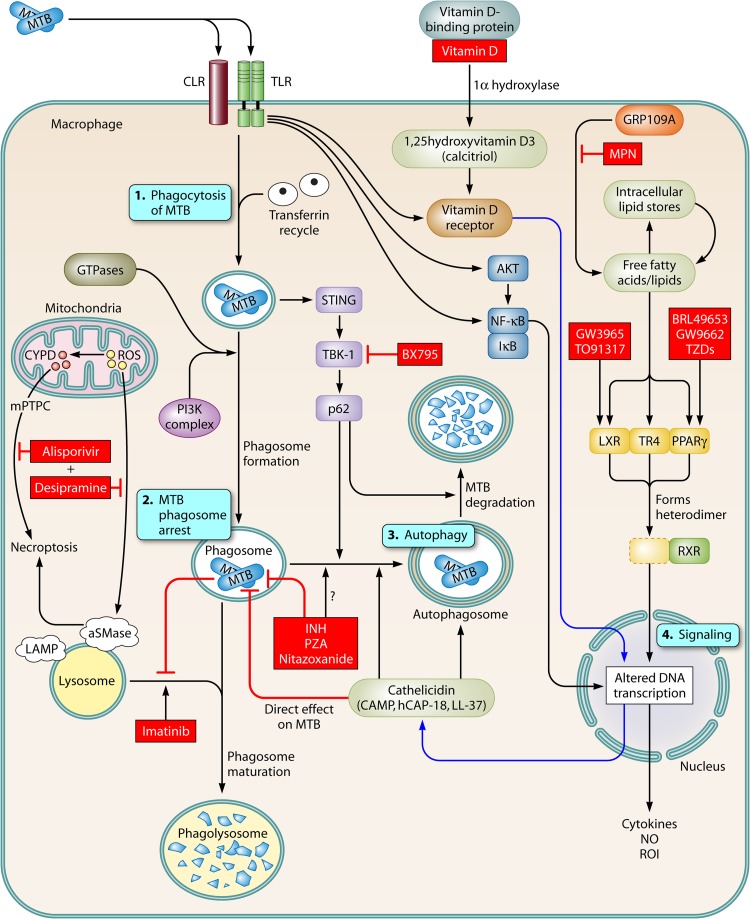
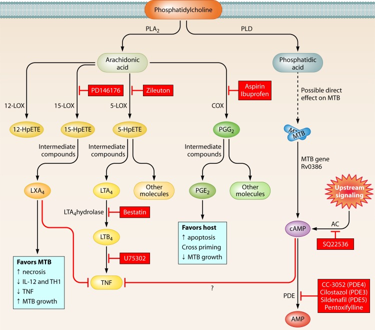
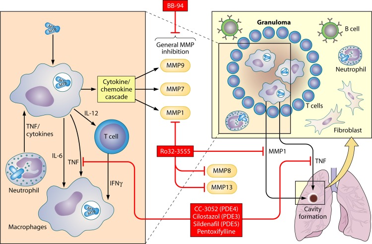
Treatment of tuberculosis (TB) remains challenging, with lengthy treatment durations and complex drug regimens that are toxic and difficult to administer. Similar to the vast majority of antibiotics, drugs for Mycobacterium tuberculosis are directed against microbial targets. Although more effective drugs that target the bacterium may lead to faster cure of patients, it is possible that a biological limit will be reached that can be overcome only by adopting a fundamentally new treatment approach. TB regimens might be improved by including agents that target host pathways. Recent work on host-pathogen interactions, host immunity, and host-directed interventions suggests that supplementing anti-TB therapy with host modulators may lead to shorter treatment times, a reduction in lung damage caused by the disease, and a lower risk of relapse or reinfection. We undertook this review to identify molecular pathways of the host that may be amenable to modulation by small molecules for the treatment of TB. Although several approaches to augmenting standard TB treatment have been proposed, only a few have been explored in detail or advanced to preclinical and clinical studies. Our review focuses on molecular targets and inhibitory small molecules that function within the macrophage or other myeloid cells, on host inflammatory pathways, or at the level of TB-induced lung pathology.


Figures

Fig 1
Fig 1
HDTs within the macrophage. Upon infection of a macrophage by M. tuberculosis, several pathways that may serve as targets for host-directed therapeutics are activated. 1. After binding and uptake of M. tuberculosis (MTB) by macrophages through innate immune receptors (e.g., C-type lectin receptors [CLRs] and Toll-like receptors [TLRs]), the bacilli are taken into a macrophage and contained in phagosomes. Several signaling pathways and molecules, including Rab proteins, IRGM1, and phosphatidylinositol 3-kinase (PI3K), promote maturation of phagosomes and fusion with lysosomes. 2. M. tuberculosis arrests the development of phagolysosomes, preventing their acidification and enabling intracellular survival of M. tuberculosis. 3. Autophagy pathways can be stimulated by M. tuberculosis or other conditions, which leads to autophagosome formation and control of M. tuberculosis growth. 4. Several pathways mediate activation of signaling molecules. TLRs activate key elements of the signaling processes, including AKT, NF-κB, and components of the vitamin D pathway. The lipid-sensing nuclear receptors, LXR, TR4, and PPARγ, bind with RXR to modulate gene expression. HDT targets described in the text are marked in boxes with red shading.
Fig 2
Fig 2
Eicosanoid pathway and regulation of inflammation and HDTs. Phosphatidylcholine in the plasma membrane is broken down by phospholipase A2 into arachidonic acid (AA) and then converted into several eicosanoids by cyclooxygenase (COX), lipoxygenase (LOX), or cytochrome P450 enzymes (not shown). 15-LOX leads to LXA4 production, which promotes cell necrosis and facilitates M. tuberculosis replication. COX enzymes lead to PGE2, which is associated with increased apoptosis and restricted M. tuberculosis growth. However, inhibition of COX enzymes with NSAIDs can lead to improved outcomes for the host. Host- or M. tuberculosis-derived adenylate cyclases also lead to increased cAMP, which is broken down by phosphodiesterases and may modulate TNF levels. HDT targets described in the text are marked in boxes with red shading.
Fig 3
Fig 3
Pulmonary immune response to M. tuberculosis infection and HDTs. M. tuberculosis-infected macrophages and dendritic cells secrete cytokines such as IL-6, IL-10, IL-12, and TNF. This cytokine response results in recruitment of macrophages, priming and differentiation of T cells, and formation of a granuloma. The nature of the host response determines M. tuberculosis replication, lung pathology, and cavity formation. HDT targets described in the text are marked in boxes with red shading.

References

    1. Berrington WR, Hawn TR. 2007. Mycobacterium tuberculosis, macrophages, and the innate immune response: does common variation matter? Immunological Rev. 219:167–186 - PMC - PubMed
    1. Ishikawa E, Ishikawa T, Morita YS, Toyonaga K, Yamada H, Takeuchi O, Kinoshita T, Akira S, Yoshikai Y, Yamasaki S. 2009. Direct recognition of the mycobacterial glycolipid, trehalose dimycolate, by C-type lectin Mincle. J. Exp. Med. 206:2879–2888 - PMC - PubMed
    1. Liu PT, Modlin RL. 2008. Human macrophage host defense against Mycobacterium tuberculosis. Curr. Opin. Immunol. 20:371–376 - PubMed
    1. Manzanillo PS, Shiloh MU, Portnoy DA, Cox JS. 2012. Mycobacterium tuberculosis activates the DNA-dependent cytosolic surveillance pathway within macrophages. Cell Host Microbe 11:469–480 - PMC - PubMed
    1. Russell DG. 2011. Mycobacterium tuberculosis and the intimate discourse of a chronic infection. Immunol. Rev. 240:252–268 - PMC - PubMed

Publication types

MeSH terms

Substances