Review
. 2014 Jan;10(1):27-36.
doi: 10.1038/nrneurol.2013.247. Epub 2013 Dec 3.

Primary cilia in neurodevelopmental disorders

Affiliations
Review

Primary cilia in neurodevelopmental disorders

Enza Maria Valente et al. Nat Rev Neurol. 2014 Jan.

Abstract

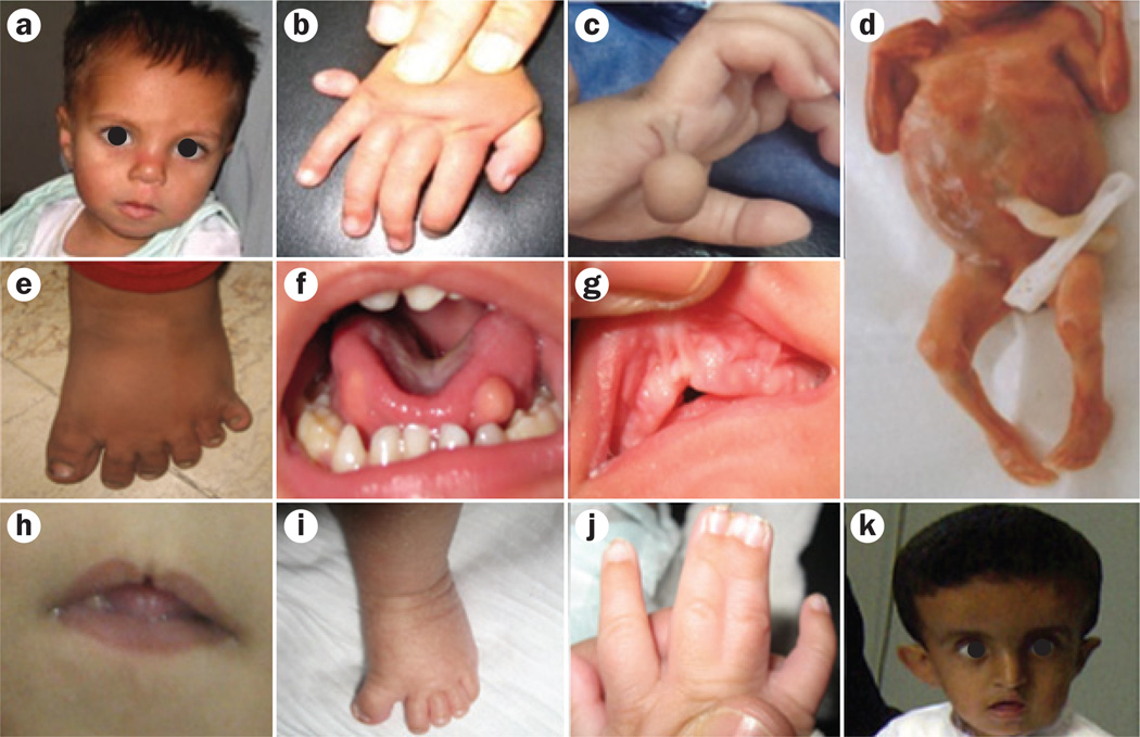
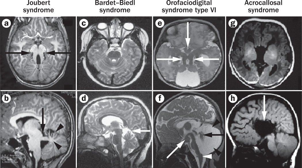
Primary cilia are generally solitary organelles that emanate from the surface of almost all vertebrate cell types. Until recently, details regarding the function of these structures were lacking; however, extensive evidence now suggests that primary cilia have critical roles in sensing the extracellular environment, and in coordinating developmental and homeostatic signalling pathways. Furthermore, disruption of these functions seems to underlie a diverse spectrum of disorders, known as primary ciliopathies. These disorders are characterized by wide-ranging clinical and genetic heterogeneity, but with substantial overlap among distinct conditions. Indeed, ciliopathies are associated with a large variety of manifestations that often include distinctive neurological findings. Herein, we review neurological features associated with primary ciliopathies, highlight genotype-phenotype correlations, and discuss potential mechanisms underlying these findings.


Figures

Figure 1
Figure 1. Structure and organization of the primary cilium
The primary cilium is a hair-like structure that protrudes from the cell surface. Microtubules form the core structure of the cilium, the axoneme. Protein cargo is transported up and down the cilium via anterograde and retrograde IFT mediated by kinesin and dynein motor proteins, respectively, which travel along the axoneme. Several proteins implicated in Joubert syndrome and Meckel syndrome form a large complex at the transition zone, which is involved in the regulation of ciliogenesis and in the control of the traffic of specific molecules into and out of the cilium. Proteins mutated in BBS cluster in the BBSome complex. This complex is relevant for ciliogenesis and regulates the correct assembly and functioning of the IFT machinery. Abbreviations: BBS, Bardet–Biedl syndrome; IFT, intraflagellar transport.
Figure 2
Figure 2. Typical clinical features in ciliopathies with CNS involvement
a | Facial dysmorphisms in JS, including prominent forehead, high and rounded eyebrows, upturned nose and anteverted nostrils. b,c | Postaxial polydactyly type B (digitus minimus) in patients with JS. d | Abdominal protrusion in a medically aborted fetus with Meckel syndrome; on autopsy, the swelling was found to be due to cystic and enlarged kidneys. e | Postaxial polydactyly type A of the foot in Bardet–Biedl syndrome. f | Hamartomatous mass of the tongue in OFD6. g | Hyperplasia of the frenula in a patient with OFD6. h | Midline cleft of the upper lip in OFD6. i | Polysyndactyly in a fibularly bent hallux in OFD6. j | Mesoaxial polysyndactyly in a patient with OFD6. k | Characteristic facies in acrocallosal syndrome. Abbreviations: JS, Joubert syndrome; OFD6, orofaciodigital syndrome type VI. Written consent for publication was obtained from the parents of the children shown in panels a and k.
Figure 3
Figure 3. Common MRI findings in ciliopathies with CNS involvement
Axial (upper panels) and midline sagittal (lower panels) brain MRI scans. a,b | Joubert syndrome. Molar tooth sign (arrows in a), with horizontally oriented and thickened superior cerebellar peduncles (arrows in a and b), and hypoplasia and dysplasia of the cerebellar vermis (arrowheads in b). c,d | Bardet–Biedl syndrome. Minimally enlarged interfoliar spaces in the superior vermis (arrow in d), with otherwise normal size and morphology of the brainstem, vermis and cerebellar hemispheres. e,f | Orofaciodigital syndrome type VI. Molar tooth sign with thickened and elongated superior cerebellar peduncles (horizontal arrows in e), hypoplasia and dysplasia of the cerebellar vermis (black arrow in f) and both cerebellar hemispheres, enlarged fourth ventricle and posterior fossa (black arrowheads in f), hypothalamic hamartoma (vertical arrow in e, white arrow in f), elongated midbrain with thin pons (not shown), and abnormal prominence of the posterior lower brainstem (white arrowhead in f). g,h | Acrocallosal syndrome. Agenesis of corpus callosum (arrow in h) with secondary abnormal configuration of the lateral ventricles; brainstem and cerebellum seem morphologically normal. Panels e and f reprinted with permission from the American Society of Neuroradiology © Poretti, A. et al. Am. J. Neuroradiol. 29, 1090–1091 (2008).

References

    1. Goetz SC, Anderson KV. The primary cilium: a signalling centre during vertebrate development. Nat. Rev. Genet. 2010;11:331–344. - PMC - PubMed
    1. Hildebrandt F, Benzing T, Katsanis N. Ciliopathies. N. Engl. J. Med. 2011;364:1533–1543. - PMC - PubMed
    1. Iannicelli M, et al. Novel TMEM67 mutations and genotype-phenotype correlates in meckelin-related ciliopathies. Hum. Mutat. 2010;31:E1319–E1331. - PMC - PubMed
    1. Zaki MS, Sattar S, Massoudi RA, Gleeson JG. Co-occurrence of distinct ciliopathy diseases in single families suggests genetic modifiers. Am. J. Med. Genet. A. 2011;155A:3042–3049. - PMC - PubMed
    1. Zaghloul NA, Katsanis N. Functional modules, mutational load and human genetic disease. Trends Genet. 2010;26:168–176. - PMC - PubMed

Publication types

MeSH terms