. 2014 Feb;24(2):161-76.
doi: 10.1038/cr.2013.155. Epub 2013 Dec 3.

Identification of IFN-γ-producing innate B cells

Affiliations

Identification of IFN-γ-producing innate B cells

Yan Bao et al. Cell Res. 2014 Feb.

Abstract

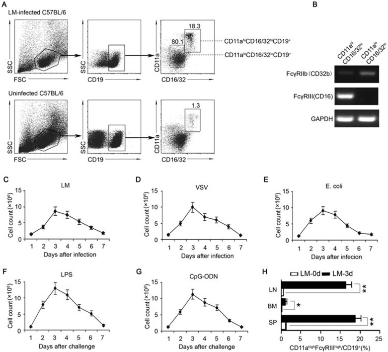
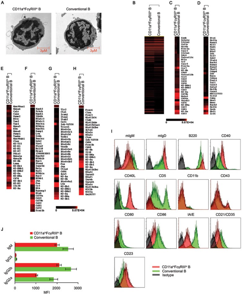
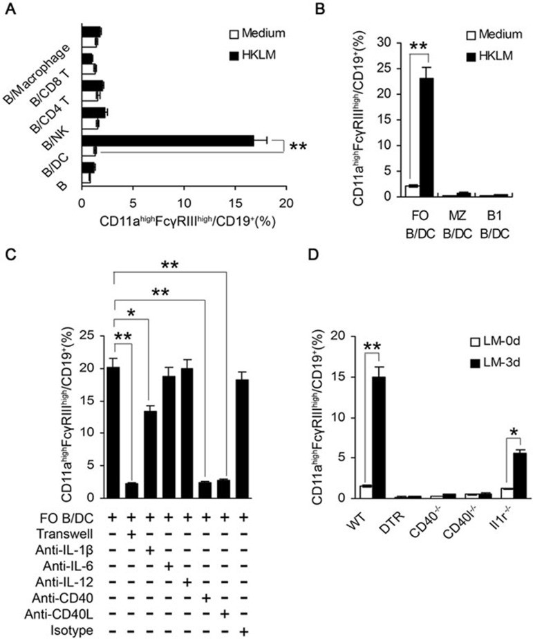
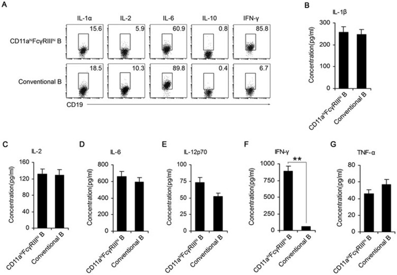
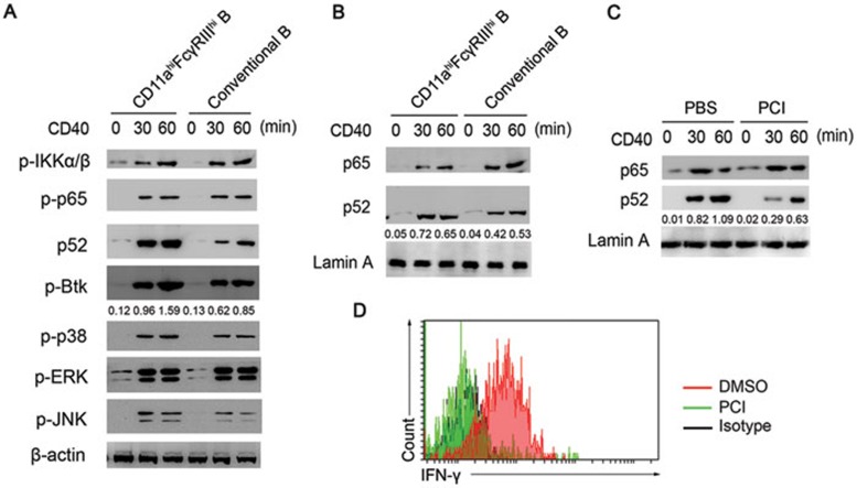
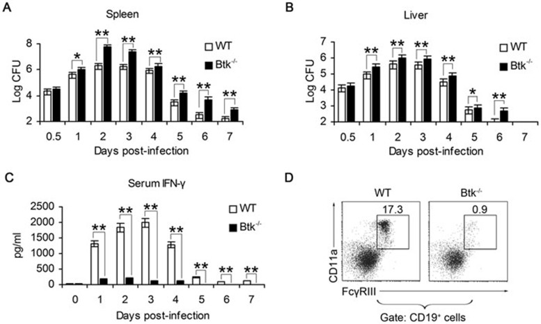
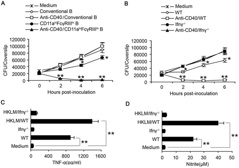
Although B cells play important roles in the humoral immune response and the regulation of adaptive immunity, B cell subpopulations with unique phenotypes, particularly those with non-classical immune functions, should be further investigated. By challenging mice with Listeria monocytogenes, Escherichia coli, vesicular stomatitis virus and Toll-like receptor ligands, we identified an inducible CD11a(hi)FcγRIII(hi) B cell subpopulation that is significantly expanded and produces high levels of IFN-γ during the early stage of the immune response. This subpopulation of B cells can promote macrophage activation via generating IFN-γ, thereby facilitating the innate immune response against intracellular bacterial infection. As this new subpopulation is of B cell origin and exhibits the phenotypic characteristics of B cells, we designated these cells as IFN-γ-producing innate B cells. Dendritic cells were essential for the inducible generation of these innate B cells from the follicular B cells via CD40L-CD40 ligation. Increased Bruton's tyrosine kinase activation was found to be responsible for the increased activation of non-canonical NF-κB pathway in these innate B cells after CD40 ligation, with the consequent induction of additional IFN-γ production. The identification of this new population of innate B cells may contribute to a better understanding of B cell functions in anti-infection immune responses and immune regulation.


Figures

Figure 1
Figure 1
Generation of CD11ahiFcγRIIIhiCD19+ cells in mice infected with pathogens or challenged with TLR ligands. Naïve C57BL/6 mice were i.p. infected with 2 × 10⁶ LM (A-C, H), 5 × 10⁶ PFU VSV (D), or 1 × 10⁶ E. coli (E). (A) Splenocytes were isolated on day 3 post-infection, and the percentage of CD11ahiFcγRIIIhiCD19+ cells in the CD19+ B cells was analyzed. (B) The FcγRIIb and FcγRIII mRNA expression of splenic CD11ahiFcγRIIIhiCD19+ or CD11aloFcγRIIIloCD19+ cells was assessed by RT-PCR. The transcript of the mouse GAPDH gene was used as an amplification control. (C-E) The number of CD11ahiFcγRIIIhi B cells in 108 splenocytes was examined within 7 days after infection with LM (C), VSV (D), and E. coli (E). (F, G) Naïve C57BL/6 mice were i.p. injected with LPS (0.5 mg/kg weight) (F) or CpG-ODN (2.5 mg/kg weight) (G). The numbers of CD11ahiFcγRIIIhi B cells in 108 splenocytes were dynamically examined within 7 days after the challenge. (H) The percentages of CD11ahiFcγRIIIhi B cells in the CD19+ B cells in the lymph nodes (LN), spleens (SP), and BM were analyzed on day 0 or day 3 post-LM infection. Data shown represent the mean ± SD. **P< 0.01, *P< 0.05.
Figure 2
Figure 2
Morphological and gene signature characteristics of the CD11ahiFcγRIIIhi B cells. C57BL/6 mice were infected with 2 × 10⁶ LM. CD11ahiFcγRIIIhi B cells and CD11aloFcγRIII conventional B cells were sorted from the splenocytes of the infected mice 3 days later. (A) Electron microscopic observation of CD11ahiFcγRIIIhi and conventional B cells. (B) Unsupervised clustering analysis of differentially expressed genes of the CD11ahiFcγRIIIhi and conventional B cells based on microarray data. Red and black correspond to high and low expression levels, respectively. (C, D) Heat-map of clustering analysis of differentially expressed cellular antigens and transcription factors in the CD11ahiFcγRIIIhi and conventional B cells. The gene symbols are listed. (E-H) Highly expressed genes in the CD11ahiFcγRIIIhi B cells were subjected to a cluster analysis with regard to the antigen processing and presentation pathway, mmu04612 (E), B cell receptor signaling pathway, mmu04662 (F), rheumatoid arthritis, mmu05323 (G), and systemic lupus erythematosus, mmu05322 (H), based on the KEGG database. The gene symbols are listed. (I) The surface markers of CD11ahiFcγRIIIhi and conventional B cells. (J) Secretion of immunoglobulins by CD11ahiFcγRIIIhi and conventional B cells after stimulation with 1 μg/ml LPS for 24 h. Data shown represent the mean ± SD. *P < 0.05.
Figure 3
Figure 3
Cellular and molecular mechanisms for the inducible generation of CD11ahiFcγRIIIhi B cells. (A) NK cells, DC, macrophages, CD4+ T cells, and CD8+ T cells were sorted from C57BL/6 mice and then co-cultured with CD19+B cells from naïve C57BL/6 mice (1:1) in the presence of HKLM (108/ml). The proportions of CD11ahiFcγRIIIhicells in the CD19+B cells were analyzed 48 h later. (B) Splenic FO (CD93CD21loCD23hi), MZ (CD93CD21hiCD23lo), and B1 (B220+CD5+) B cells were purified and co-cultured with DCs. HKLM was added in the co-culture system. The percentages of CD11ahiFcγRIIIhicells in the CD19+ B cells were assessed 48 h later. (C) DCs and FO B cells from WT mice were co-cultured in the presence of HKLM. Anti-IL-1β (5 μg/ml), anti-IL-6 (5 μg/ml), anti-IL-12 (5 μg/ml), anti-CD40 (5 μg/ml), or anti-CD40L (5 μg/ml) was added or a 0.4-μm transwell system was used, as indicated. The expression of CD11a and FcγRIII on B cells was examined after 48 h. (D) CD11c-DTR mice were injected with diphtheria toxin (DT) (100 ng) for the depletion of conventional DCs. The CD11ahiFcγRIIIhi cell population in splenic B cells was determined in WT, DC-depleted (DTR), Cd40−/−, Cd40l−/− and Il1r−/− mice 3 days after LM infection. Data shown represent the mean ± SD of triplicate experiments. *P< 0.05, **P < 0.01.
Figure 4
Figure 4
CD11ahiFcγRIIIhi B cells produce a high level of IFN-γ in response to CD40 ligation. C57BL/6 mice were infected with 2 × 10⁶ LM. (A) The intracellular expression of IL-1α, IL-2, IL-6, IL-10 and IFN-γ in CD11ahiFcγRIIIhi and conventional B cells was assayed on day 3 post-infection. (B-G) Splenic CD11ahiFcγRIIIhi and conventional B cells were sorted on day 3 post-infection. IL-1β, IL-2, IL-6, IL-12p70, IFN-γ, or TNF-α secretion by CD11ahiFcγRIIIhi and conventional B cells was detected by ELISA. Data shown represent the mean ± SD of triplicate experiments. *P< 0.05, **P < 0.01.
Figure 5
Figure 5
Increased activation of the Btk and non-canonical NF-κB pathways is responsible for the increased IFN-γ production in CD40-triggered CD11ahiFcγRIIIhi B cells. (A, B) Signaling pathways in the CD11ahiFcγRIIIhi and conventional B cells stimulated with activating anti-CD40, with actin (A) and lamin A (B) as loading controls. The data are representative of three independent experiments with similar results. The numbers below the lanes (top) indicate Btk (A) and p52 (B, C) band densities, presented relative to the β-actin (A) and lamin A (B, C) expression in the same lane (below). (C) Nuclear translocation of p65 and p52 in CD11ahiFcγRIIIhi B cells pretreated with the Btk inhibitor PCI-32765 (5 nM) for 60 min. (D) Intracellular IFN-γ expression in CD11ahiFcγRIIIhi B cells pretreated with neutralizing CD11a mAbs or the Btk inhibitor PCI-32765.
Figure 6
Figure 6
Btk−/− mice with impaired CD11ahiFcγRIIIhi B cell generation are more susceptible to LM infection. WT and Btk−/− mice were infected with LM. CFUs in the spleen (A) and liver (B) and serum IFN-γ (C) were analyzed at the indicated time. (D) The CD11ahiFcγRIIIhi cell proportion in splenic B cells was determined in WT and Btk−/− mice on day 3 post-LM infection. Data shown represent the mean ± SD of triplicate experiments. *P < 0.05, **P < 0.01.
Figure 7
Figure 7
CD11ahiFcγRIIIhi B cells enhance the resistance of macrophages to LM infection through IFN-γ. (A, B) BMDMs from WT mice were infected with LM. (A) LM-infected BMDMs were co-cultured with CD11ahiFcγRIIIhi or conventional B cells from WT mice with or without anti-CD40 pretreatment. (B) LM-infected BMDMs were co-cultured with CD11ahiFcγRIIIhi B cells from WT or Ifnγ−/− mice with or without anti-CD40 pretreatment. (A, B) CFUs/coverslip (mean ± SD) were determined at 0, 2, 4, and 6 h post-infection. (C, D) BMDMs were co-cultured with anti-CD40-pretreated CD11ahiFcγRIIIhi B cells from WT or Ifnγ−/− mice in the presence or absence of HKLM. After 48 h, the TNF-α (C) and nitrite (D) levels in the supernatants were examined. *P< 0.05, **P < 0.01.
Figure 8
Figure 8
Adoptive transfer of CD11ahiFcγRIIIhi B cells promotes the innate defense of Btk−/− mice against LM infection. CD11ahiFcγRIIIhi and conventional B cells were purified from the spleen of WT or Ifnγ−/− mice on day 3 after infection with LM using Dako MoFloTM XDP; the cells were then transferred intravenously into Btk−/− mice infected with LM. CFUs in the spleen (A) and liver (B), and serum IFN-γ (C) were examined at the indicated time. Data shown represent the mean ± SD of triplicate experiments. *P < 0.05, **P < 0.01.

Comment in

  • Innate IFNγ-producing B cells.
    Ballesteros-Tato A, Stone SL, Lund FE. Ballesteros-Tato A, et al. Cell Res. 2014 Feb;24(2):135-6. doi: 10.1038/cr.2013.163. Epub 2013 Dec 17. Cell Res. 2014. PMID: 24343577 Free PMC article.

References

    1. Baumgarth N. The double life of a B-1 cell: self-reactivity selects for protective effector functions. Nat Rev Immunol. 2011;11:34–46. - PubMed
    1. Lund FE, Randall TD. Effector and regulatory B cells: modulators of CD4+ T cell immunity. Nat Rev Immunol. 2010;10:236–247. - PMC - PubMed
    1. Lund FE. Cytokine-producing B lymphocytes-key regulators of immunity. Curr Opin Immunol. 2008;20:332–338. - PMC - PubMed
    1. Baumgarth N, Herman OC, Jager GC, et al. B-1 and B-2 cell-derived immunoglobulin M antibodies are nonredundant components of the protective response to influenza virus infection. J Exp Med. 2000;192:271–280. - PMC - PubMed
    1. Mauri C, Ehrenstein MR. The 'short' history of regulatory B cells. Trends Immunol. 2008;29:34–40. - PubMed

Publication types

MeSH terms