. 2014 Jan 17;289(3):1592-603.
doi: 10.1074/jbc.M113.507228. Epub 2013 Dec 2.

Connexin43 reduces melanoma growth within a keratinocyte microenvironment and during tumorigenesis in vivo

Affiliations

Connexin43 reduces melanoma growth within a keratinocyte microenvironment and during tumorigenesis in vivo

Mark J Ableser et al. J Biol Chem. .

Abstract

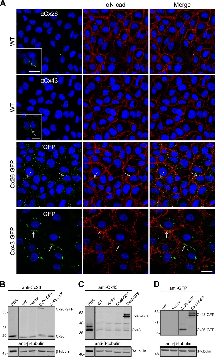
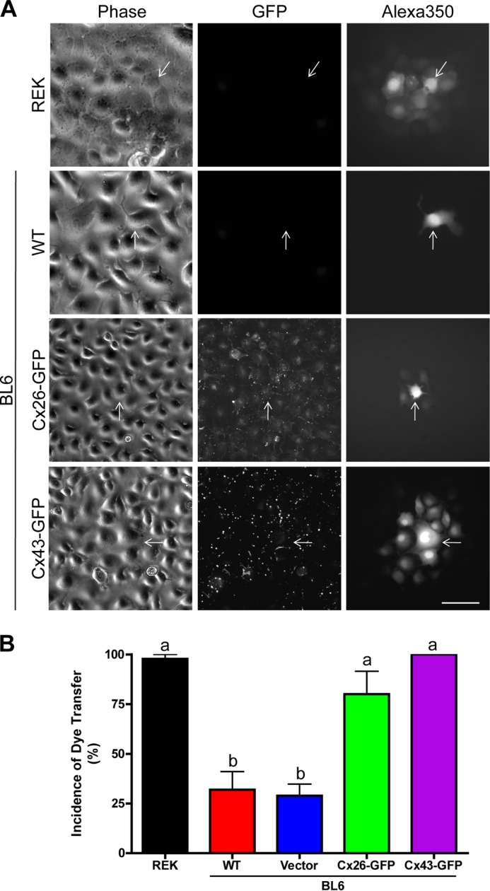
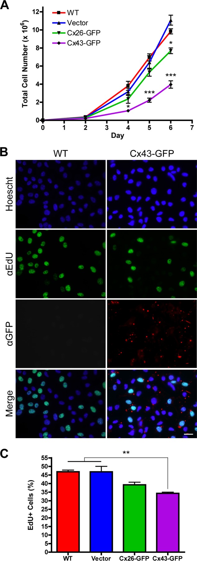
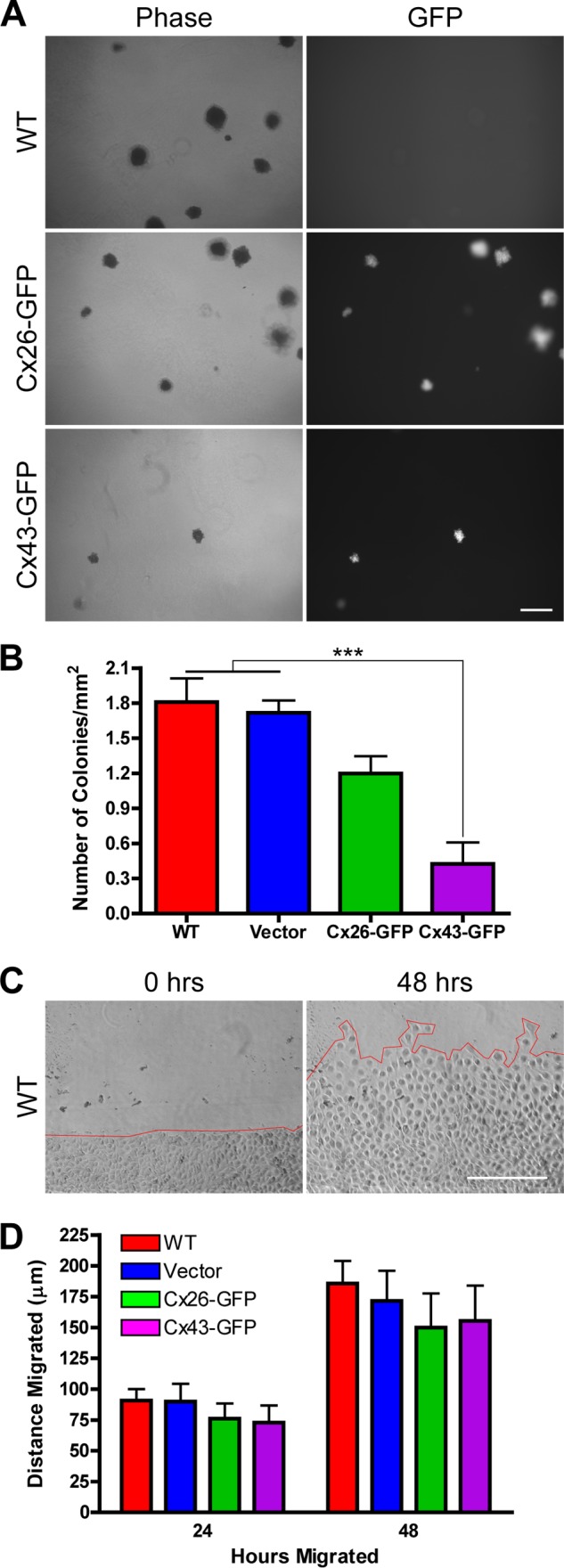
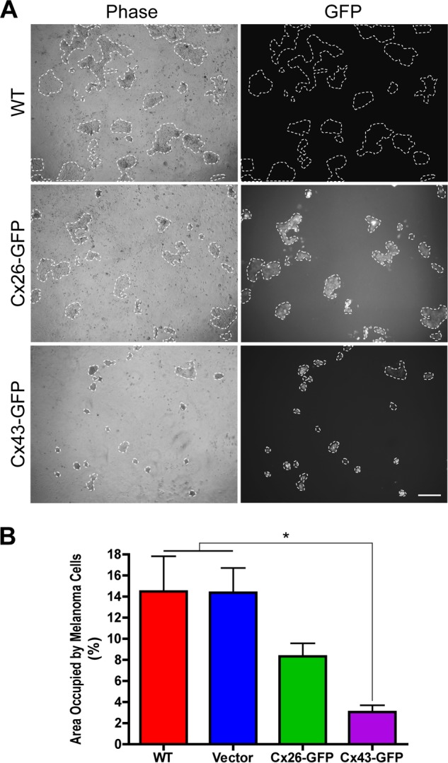
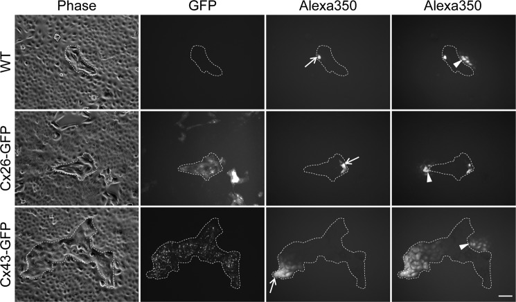
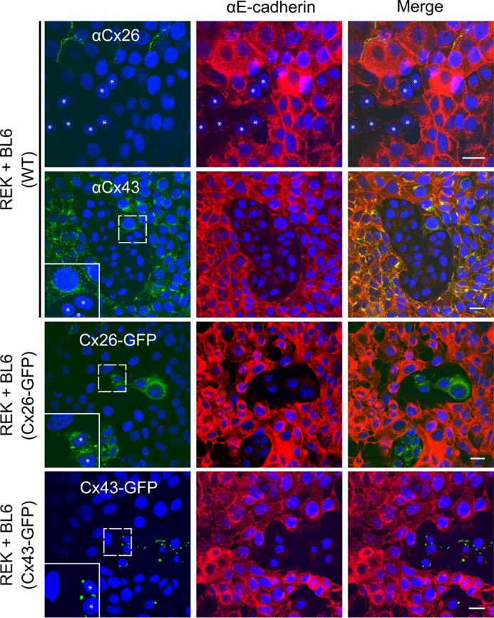
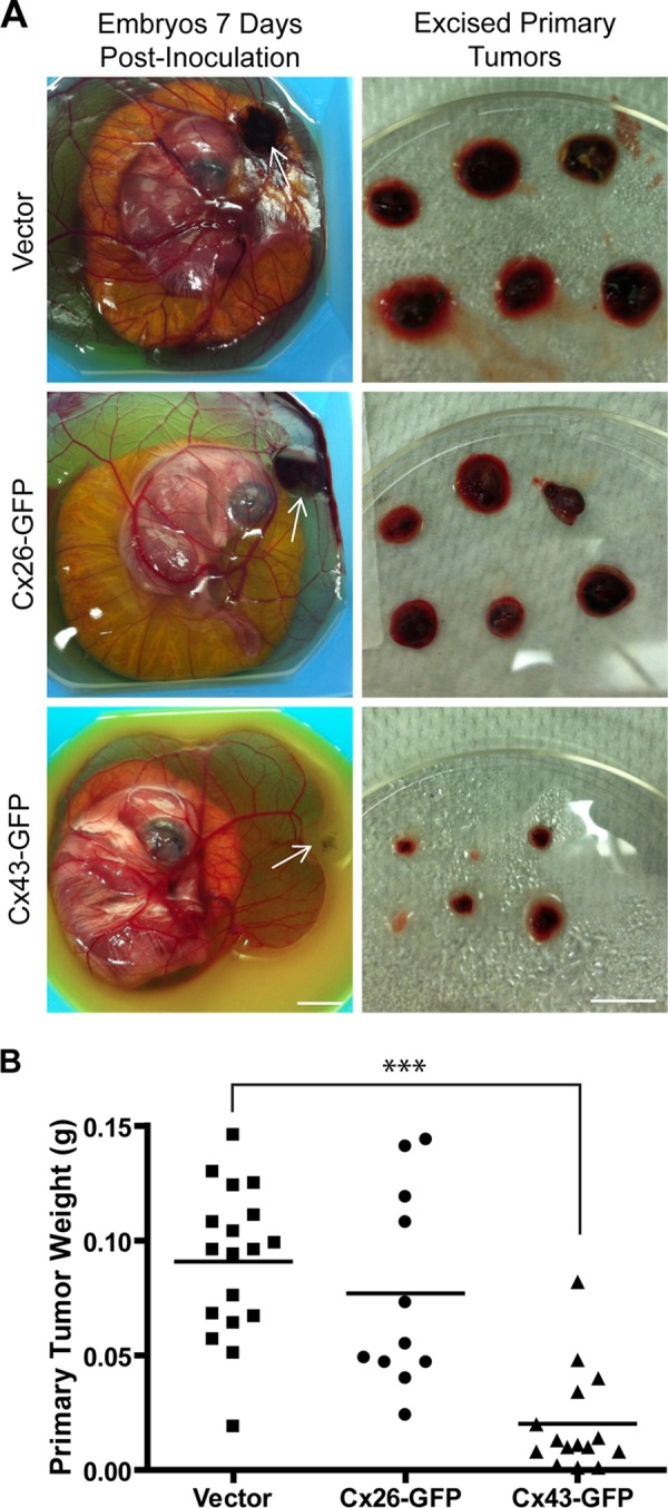
Connexins (Cx) have been identified as tumor suppressors or enhancers, a distinction that appears to be dependent on the type and stage of disease. However, the role of connexins in melanoma tumorigenesis and their status during cancer onset and progression remain controversial and unclear. Here, we show that the aggressive B16-BL6 mouse melanoma cell line expresses low basal levels of Cx26 and Cx43, rendering them gap junctional intercellular communication-deficient as elucidated by immunofluorescence, Western blotting, and dye transfer studies. Following ectopic expression of green fluorescent protein-tagged Cx26 and Cx43 in these connexin-deficient melanomas, punctate gap junction-like plaques were evident at sites of cell-cell apposition, and the incidence of dye transfer was significantly increased similar to connexin-rich keratinocytes. We found that the expression of Cx43, but not Cx26, significantly reduced cellular proliferation and anchorage-independent growth from control melanomas, whereas migration was unaffected. Additionally, melanomas expressing Cx43 displayed significantly reduced growth within the in situ-like microenvironment of keratinocytes, despite a lack of heterocellular gap junctional intercellular communication between the two cell types. Furthermore, when grown in vivo in the chicken chorioallantoic membrane, primary tumors derived from Cx43-expressing melanomas were significantly smaller than controls, whereas Cx26-expressing melanomas produced tumors similar to controls. Collectively, these results suggest that Cx43, and not Cx26, can act as a tumor suppressor during melanoma tumorigenesis.

Keywords: Connexin; Gap Junctions; Keratinocytes; Melanoma; Tumor Suppressor Gene.


Figures

FIGURE 1.
FIGURE 1.
BL6 mouse melanomas express low levels of Cx26 and Cx43. A, immunofluorescence revealed that WT BL6 cells endogenously express low levels of Cx26 and Cx43 compared with REKs, which display typical gap junctional plaques at sites of cell-cell apposition (inserts). B and C, Western blots confirmed the low levels of Cx26 and Cx43 in melanomas compared with lysates collected from over-confluent and confluent REK cultures, respectively. Following ectopic expression of Cx26-GFP or Cx43-GFP, punctate gap junction-like plaques were evident at the cell surface as denoted by the cell adhesion molecule N-cadherin (red) (A, arrows), and the expression of GFP-tagged connexins was readily detected by Western blots immunolabeled for Cx26 (B) or Cx43 (C). D, additionally, both fusion proteins were expressed at similar levels as detected by Western blots immunolabeled for GFP. β-Tubulin was used as a loading control. The vector control represents cells transfected with a construct that did not encode connexins. Lanes separated by vertical lines were run on the same blot but spliced together. Lanes loaded with REK lysates were run as independent experiments. Blue, nuclei; bars, 20 μm.
FIGURE 2.
FIGURE 2.
Ectopic connexin expression significantly increases GJIC. A, Alexa350 dye transfer studies revealed that wild-type and control melanomas were poorly coupled, because dye rarely spread from the microinjected cell (arrows) to neighboring cells. B, following ectopic expression of Cx26-GFP or Cx43-GFP, the incidence of dye transfer was significantly increased to 80 and 100%, respectively, statistically similar to Cx43-rich REK controls (n = 45, p < 0.05). Phase contrast images depict cellular morphology prior to microinjection, whereas the GFP fluorescence denotes the expression of ectopic connexins. Letters depict statistical significance among the groups. Bar, 40 μm.
FIGURE 3.
FIGURE 3.
Expression of Cx43 significantly reduces total cell number and cell proliferation. A, 6-day growth curves revealed that Cx43-GFP-expressing melanomas displayed significantly reduced total cell number as early as day 4 (n = 8; *, p < 0.05), which was further reduced on days 5 and 6 (n = 8; ***, p < 0.001). Cx26-GFP-expressing melanomas displayed a slight reduction in total cell number only on day 6 (n = 8, *, p < 0.05). B, proliferation at day 4 was assessed using an EdU assay, which labels nuclei passing through the S phase of the cell cycle, where the proportion of EdU-labeled nuclei (green) to total nuclei labeled with Hoescht (blue) was determined. Cells were counterstained with an anti-GFP antibody (red) because the normal fluorescence of GFP-tagged Cx43 was quenched by the EdU signal. C, expression of Cx43 significantly reduced the percentage of EdU-positive cells by ∼25% in comparison to the controls (n = 40; **, p < 0.01). Bar, 20 μm.
FIGURE 4.
FIGURE 4.
Cx43 expression significantly reduces anchorage-independent growth, whereas cell migration remains unaffected. A, when grown in soft agar suspension for 10 days, cell colonies were evident under phase contrast, and those formed from Cx-expressing melanomas were GFP-positive. B, the number of colonies formed was significantly reduced in melanomas expressing Cx43-GFP by ∼4.5-fold in comparison to controls (n = 240; ***, p < 0.001), whereas Cx26-GFP expression did not significantly alter the number of cell colonies (n = 240, p = ns). C, using a scratch assay, the migratory ability of Cx-deficient and Cx-expressing melanomas was assessed. Cells were grown to confluence, pulsed with 25 μg/ml of Mitomycin C for 30 min, scratched (red line at 0 h), and supplemented with serum-free medium. D, the distance migrated from the initial scratch edge to the leading edge after 24 and 48 h (C, red line at 48 h) was not statistically different between Cx-deficient and Cx-expressing melanomas (n = 20, p = ns). Bars, 200 μm.
FIGURE 5.
FIGURE 5.
Expression of Cx43 significantly reduces melanoma growth within a keratinocyte-based microenvironment. A, melanomas were co-cultured with keratinocytes at a 1:5 ratio and allowed to grow for 6 days. Under phase contrast imaging, melanoma clusters were identifiable (dashed lines), and their melanoma phenotype was confirmed in engineered cell lines because of the expression of GFP. B, the area of all melanoma clusters for a given field was measured, and Cx43-expressing melanomas were found to occupy ∼4.5-fold less area than controls (n = 40; *, p < 0.05). Bar, 200 μm.
FIGURE 6.
FIGURE 6.
Ectopic connexin expression in melanomas does not establish heterocellular GJIC with keratinocytes. Melanomas were co-cultured with keratinocytes at a 1:10 ratio, respectively. Under phase contrast, melanoma clusters were identified (dashed lines), and their melanoma phenotypes were confirmed, where possible, by the expression of GFP. In all cases, injected melanomas (arrows) failed to pass Alexa350 to adjacent keratinocytes, and only Cx-expressing melanomas exhibited sufficient dye transfer among each other (n = 18). Similarly, keratinocytes injected with Alexa350 (arrowheads) were highly coupled with neighboring keratinocytes, but in all cases, failed to pass dye to adjacent melanomas (n = 18). Bar, 50 μm.
FIGURE 7.
FIGURE 7.
Cx26 or Cx43 do not act to form melanoma/keratinocyte heterocellular gap junctions. REKs were co-cultured with WT BL6 cells or cells engineered to express Cx26-GFP or Cx43-GFP. Heterocellular cultures were double immunolabeled for Cx26 or Cx43 and E-cadherin or immunolabeled for E-cadherin only. In all cases, control or connexin-expressing BL6 cells did not exhibit clear evidence of Cx43 or Cx26 gap junction plaques at interfaces with E-cadherin-positive REKs even when examined under high magnification (boxed insets). Asterisks highlight some BL6 cells. Inset images were magnified 2×. Bars, 20 μm.
FIGURE 8.
FIGURE 8.
Cx43 expression significantly reduces primary tumor growth in vivo. A, 300,000 control or Cx-expressing melanomas were topically applied to the CAM of 10-day-old chicken embryos. Primary melanoma tumors visible on the CAM 7 days post-inoculation (arrows) were excised, photographed, and weighed. B, control melanomas produced large, blood-filled tumors of ∼0.09 g in weight (n = 18). Cx26-GFP-expressing melanomas exhibited a slight reduction in primary tumor weight (∼0.075 g), but this was not statistically significant (n = 11, p = ns). Conversely, melanomas expressing Cx43-GFP produced significantly smaller tumors of ∼0.02 g in weight (∼4.5-fold reduction from controls) (n = 15; ***, p < 0.001). Bars, 1 cm.

References

    1. Söhl G., Willecke K. (2003) An update on connexin genes and their nomenclature in mouse and man. Cell Commun. Adhes. 10, 173–180 - PubMed
    1. Wang N., De Bock M., Decrock E., Bol M., Gadicherla A., Vinken M., Rogiers V., Bukauskas F. F., Bultynck G., Leybaert L. (2013) Paracrine signaling through plasma membrane hemichannels. Biochim. Biophys. Acta 1828, 35–50 - PMC - PubMed
    1. Richard G. (2000) Connexins. A connection with the skin. Exp. Dermatol. 9, 77–96 - PubMed
    1. Hsu M., Andl T., Li G., Meinkoth J. L., Herlyn M. (2000) Cadherin repertoire determines partner-specific gap junctional communication during melanoma progression. J. Cell Sci. 113, 1535–1542 - PubMed
    1. Masuda M., Usami S., Yamazaki K., Takumi Y., Shinkawa H., Kurashima K., Kunihiro T., Kanzaki J. (2001) Connexin 26 distribution in gap junctions between melanocytes in the human vestibular dark cell area. Anat. Rec. 262, 137–146 - PubMed

Publication types

MeSH terms