

Protective effect of metformin against cisplatin-induced ototoxicity in an auditory cell line
- PMID: 24297263
- PMCID: PMC3946136
- DOI: 10.1007/s10162-013-0431-y
Protective effect of metformin against cisplatin-induced ototoxicity in an auditory cell line
Abstract
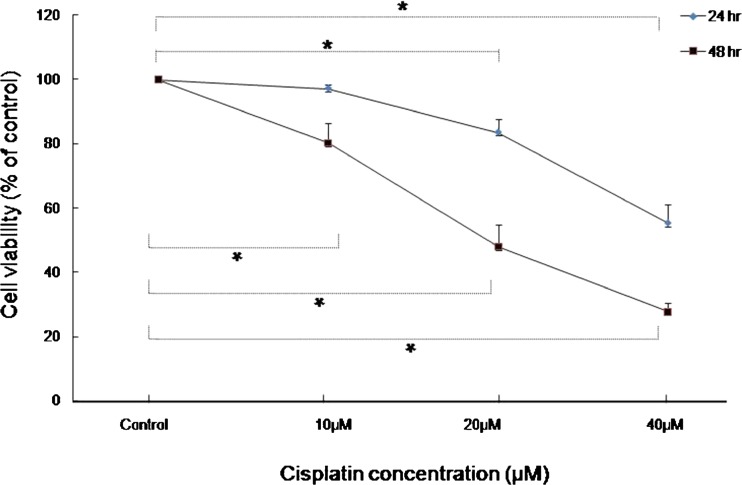
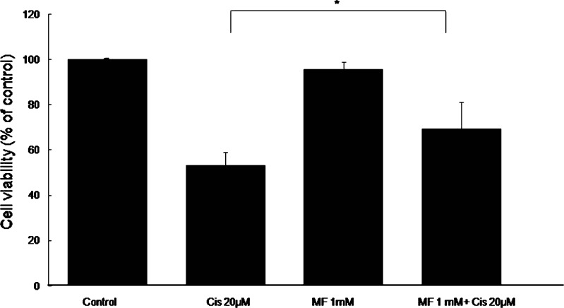
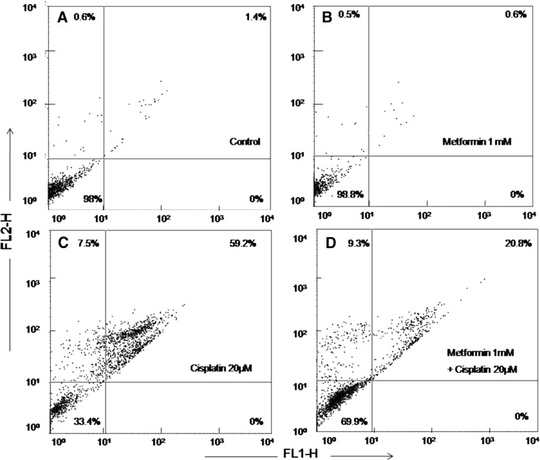
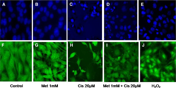
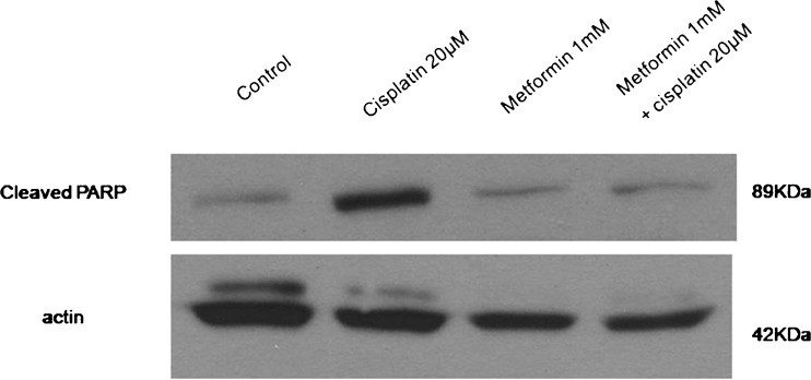
Metformin, an antidiabetic drug with potent anticancer activity, is known to prevent oxidative stress-induced cell death in several cell types through a mechanism dependent on the mitochondria. In the present study, we investigated the influence of metformin on cisplatin ototoxicity in an auditory cell line. Cell viability was determined using a 3-(4,5-dimethylthiazol-2-yl)-2,5-diphenyltetrazoliumbromide (Sigma, St. Louis, MO, USA) cell proliferation assay. Oxidative stress and apoptosis were assessed by flow cytometry analysis, Hoechst 33258 staining, reactive oxygen species (ROS) measurement, and western blotting. Intracellular calcium concentration changes were detected using calcium imaging. Pretreatment with 1 mM metformin prior to the application of 20 μM cisplatin significantly decreased the frequency of late apoptosis in HEI-OC1 cells and also significantly attenuated the cisplatin-induced increase in ROS. In addition, metformin inhibited the activation of caspase-3 and levels of poly-ADP-ribose polymerase (PARP). Pretreatment with metformin prevented the cisplatin-induced elevation in intracellular calcium concentrations. We propose that metformin protects against cisplatin-induced ototoxicity by inhibiting the increase in intracellular calcium levels, preventing apoptosis, and limiting ROS production.
Figures


Similar articles
-
Protective role of edaravone against cisplatin-induced ototoxicity in an auditory cell line.Hear Res. 2015 Dec;330(Pt A):113-8. doi: 10.1016/j.heares.2015.08.004. Epub 2015 Aug 13. Hear Res. 2015. PMID: 26278638
-
Protective Effect of Tempol against Cisplatin-Induced Ototoxicity.Int J Mol Sci. 2016 Nov 18;17(11):1931. doi: 10.3390/ijms17111931. Int J Mol Sci. 2016. PMID: 27869744 Free PMC article.
-
Protective role of antidiabetic drug metformin against gentamicin induced apoptosis in auditory cell line.Hear Res. 2011 Dec;282(1-2):92-6. doi: 10.1016/j.heares.2011.09.005. Epub 2011 Sep 28. Hear Res. 2011. PMID: 21979311
-
The effects of the antioxidant α-tocopherol succinate on cisplatin-induced ototoxicity in HEI-OC1 auditory cells.Int J Pediatr Otorhinolaryngol. 2016 Jul;86:9-14. doi: 10.1016/j.ijporl.2016.04.008. Epub 2016 Apr 12. Int J Pediatr Otorhinolaryngol. 2016. PMID: 27260571
-
[Aminoglycoside- and cisplatin-ototoxicity: from basic science to clinics].Laryngorhinootologie. 2004 May;83(5):317-23. doi: 10.1055/s-2004-814280. Laryngorhinootologie. 2004. PMID: 15143449 Review. German.
Cited by
-
Metformin as a cellular protector; a synoptic view of modern evidences.J Nephropharmacol. 2015 Jan 1;4(1):31-36. eCollection 2015. J Nephropharmacol. 2015. PMID: 28197472 Free PMC article. Review.
-
Antidiabetic Drug Metformin Protects Neuronal Cells against Quinolinic Acid-Induced Excitotoxicity by Decreasing Intracellular Calcium.Chonnam Med J. 2018 Jan;54(1):24-30. doi: 10.4068/cmj.2018.54.1.24. Epub 2018 Jan 25. Chonnam Med J. 2018. PMID: 29399562 Free PMC article.
-
Metformin in combination with cisplatin inhibits cell viability and induces apoptosis of human ovarian cancer cells by inactivating ERK 1/2.Oncol Lett. 2017 Dec;14(6):7557-7564. doi: 10.3892/ol.2017.7176. Epub 2017 Oct 12. Oncol Lett. 2017. PMID: 29344202 Free PMC article.
-
Histone deacetylase inhibition prevents cell death induced by loss of tricellular tight junction proteins in temperature-sensitive mouse cochlear cells.PLoS One. 2017 Aug 2;12(8):e0182291. doi: 10.1371/journal.pone.0182291. eCollection 2017. PLoS One. 2017. PMID: 28767685 Free PMC article.
-
Metformin Ameliorates 2.856 GHz Microwave- Radiation-Induced Reproductive Impairments in Male Rats via Inhibition of Oxidative Stress and Apoptosis.Int J Mol Sci. 2023 Jul 31;24(15):12250. doi: 10.3390/ijms241512250. Int J Mol Sci. 2023. PMID: 37569626 Free PMC article.
References
-
- Anisimov VN, Berstein LM, Egormin PA, Piskunova TS, Popovich IG, Zabezhinski MA, Kovalenko IG, Poroshina TE, Semenchenko AV, Provinciali M, Re F, Franceschi C. Effect of metformin on life span and on the development of spontaneous mammary tumors in HER-2/neu transgenic mice. Exp Gerontol. 2005;40:685–693. doi: 10.1016/j.exger.2005.07.007. - DOI - PubMed
-
- Boulikas T, Vougiouka M. Cisplatin and platinum drugs at the molecular level: review. Oncol Rep. 2003;10(6):1663–1682. - PubMed
Publication types
MeSH terms
Substances
LinkOut - more resources
Full Text Sources
Other Literature Sources
Research Materials
