

Treatment of displaced talar neck fractures using delayed procedures of plate fixation through dual approaches
- PMID: 24297608
- PMCID: PMC3890131
- DOI: 10.1007/s00264-013-2164-2
Treatment of displaced talar neck fractures using delayed procedures of plate fixation through dual approaches
Abstract
Purpose: Treatment of talar neck fractures is challenging. Various surgical approaches and fixation methods have been documented. Clinical outcomes are often dissatisfying due to inadequate reduction and fixation with high rates of complications. Obtaining satisfactory clinical outcomes with minimum complications remains a hard task for orthopaedic surgeons.
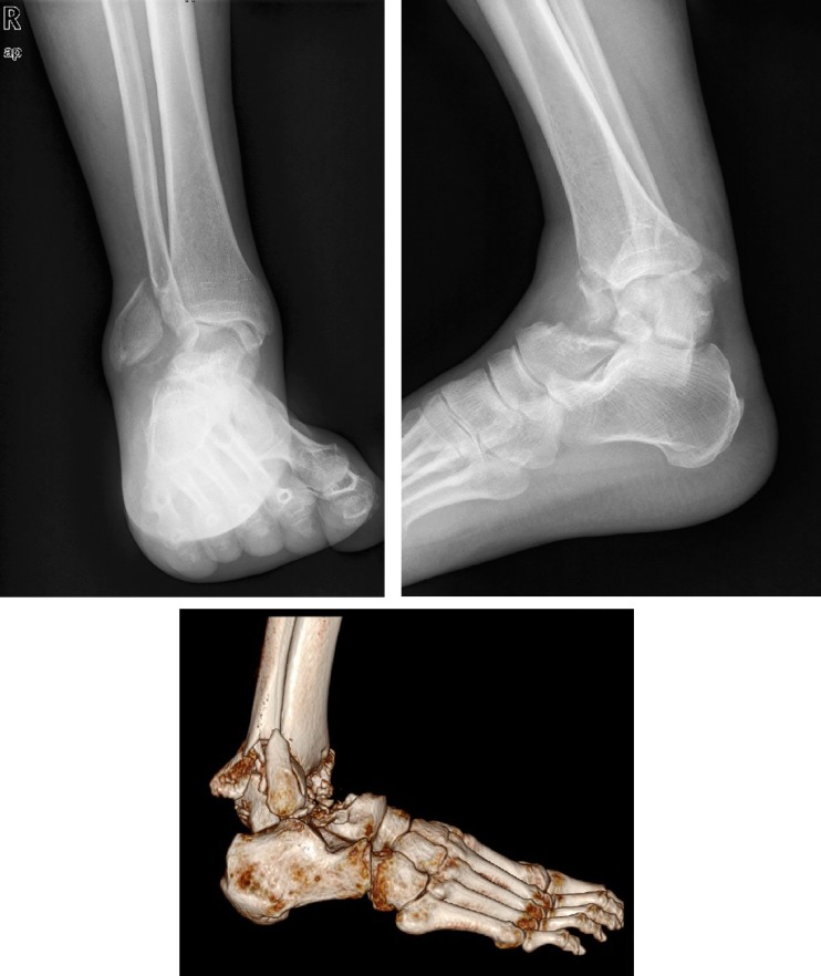
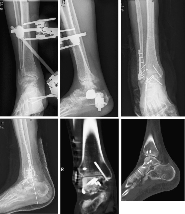
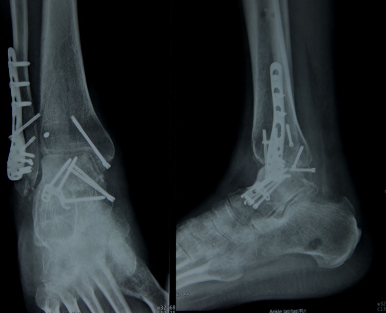
Methods: In the period from May 2007 to September 2010, a total of 31 cases with closed displaced talar neck fractures were treated surgically in our department. Injuries were classified according to the Hawkins classification modified by Canale and Kelly. Under general anaesthesia with sufficient muscle relaxation, urgent closed reduction was initiated once the patients were admitted; if the procedure failed, open reduction and provisional stabilisation with Kirschner wires through an anteromedial approach with tibiometatarsal external fixation were performed. When the soft tissue had recovered, definitive fixation was performed with plate and screws through dual approaches. The final follow-up examination included radiological analysis, clinical evaluation and functional outcomes which were carried out according to the Ankle-Hindfoot Scale of the American Orthopaedic Foot and Ankle Society (AOFAS), patient satisfaction and SF-36.
Results: Twenty-eight patients were followed up for an average of 25 months (range 18-50 months) after the injury. Only two patients had soft tissue complications, and recovery was satisfactory with conservative treatment. All of the fractures healed anatomically without malunion and nonunion, and the average union time was 14 weeks (range 12-24 weeks). Post-traumatic arthritis developed in ten cases, while six patients suffered from avascular necrosis of the talus. Secondary procedures included three cases of subtalar arthrodesis, one case of ankle arthrodesis and one case of total ankle replacement. The mean AOFAS hindfoot score was 78 (range 65-91). According to the SF-36, the average score of the physical component summary was 68 (range 59-81), and the average score of the mental component summary was 74 (range 63-85).
Conclusions: Talar neck fractures are associated with a high incidence of long-term disability and complications. Urgent reduction of the fracture-dislocation and delayed plate fixation through a dual approach when the soft tissue has recovered may minimise the complications and provide good clinical outcomes.
Figures
References
-
- García-Rey E, Sanz-Hospital FJ, Galdrán FJ, Cano-Egea JM, Alcázar LFL. Talar neck fractures: results and complications by type. Foot Ankle Surg. 2002;8:203–208. doi: 10.1046/j.1460-9584.2002.00305.x. - DOI
-
- Hawkins LG. Fractures of the neck of the talus. J Bone Joint Surg Am. 1970;52(5):991–1002. - PubMed
MeSH terms
LinkOut - more resources
Full Text Sources
Other Literature Sources
Medical
