Clinical Trial
. 2013 Dec 17;110(51):20783-8.
doi: 10.1073/pnas.1313944110. Epub 2013 Dec 2.

MuSK IgG4 autoantibodies cause myasthenia gravis by inhibiting binding between MuSK and Lrp4

Affiliations
Clinical Trial

MuSK IgG4 autoantibodies cause myasthenia gravis by inhibiting binding between MuSK and Lrp4

Maartje G Huijbers et al. Proc Natl Acad Sci U S A. .

Abstract

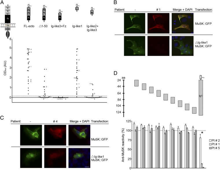
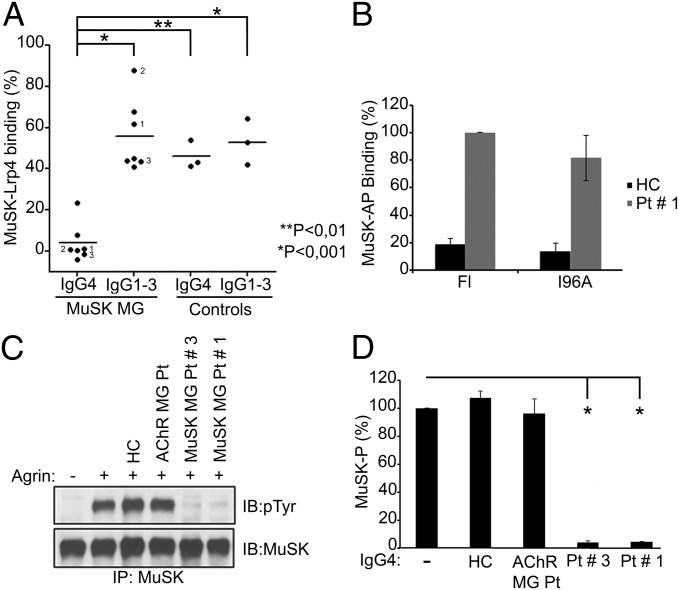
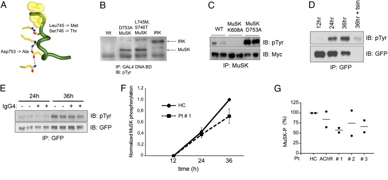
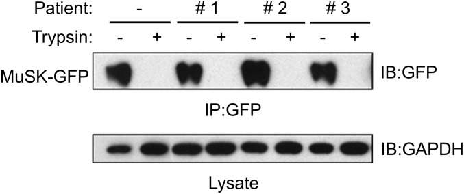
Myasthenia gravis (MG) is a severely debilitating autoimmune disease that is due to a decrease in the efficiency of synaptic transmission at neuromuscular synapses. MG is caused by antibodies against postsynaptic proteins, including (i) acetylcholine receptors, the neurotransmitter receptor, (ii) muscle-specific kinase (MuSK), a receptor tyrosine kinase essential for the formation and maintenance of neuromuscular synapses, and (iii) low-density lipoprotein receptor-related protein 4 (Lrp4), which responds to neural Agrin by binding and stimulating MuSK. Passive transfer studies in mice have shown that IgG4 antibodies from MuSK MG patients cause disease without requiring complement or other immune components, suggesting that these MuSK antibodies cause disease by directly interfering with MuSK function. Here we show that pathogenic IgG4 antibodies to MuSK bind to a structural epitope in the first Ig-like domain of MuSK, prevent binding between MuSK and Lrp4, and inhibit Agrin-stimulated MuSK phosphorylation. In contrast, these IgG4 antibodies have no direct effect on MuSK dimerization or MuSK internalization. These results provide insight into the unique pathogenesis of MuSK MG and provide clues toward development of specific treatment options.

Keywords: Dok7; Rapsyn; activation loop; insulin receptor; neuromuscular junction.


Conflict of interest statement

The authors declare no conflict of interest.

Figures

Fig. 1.
Fig. 1.
MuSK MG IgG4 antibodies bind predominantly to the first Ig-like domain in MuSK. (A) An ELISA shows that antibodies from all 25 patients bind to the extracellular region of MuSK. The predominant binding sites reside in the first Ig-like domain, because antibodies bind to this domain nearly as well as the entire extracellular region. Moreover, deletion of the N-terminal half of the first Ig-like domain substantially reduces antibody binding. Antibodies from five patients have additional reactivity to the second Ig-like domain. Data shown reflect the average binding per patient determined in three independent assays (Fig. S1). (B and C) Antibodies that bind selectively to the first Ig-like domain stain cells expressing full-length MuSK-GFP but not ∆Ig-like1-MuSK-GFP, whereas antibodies with additional reactivity bind to cells expressing either construct. (D) An ELISA shows that antibody-binding to the extracellular region of MuSK is strongly inhibited by the first Ig-like domain but poorly by 20-mer overlapping peptides from this domain.
Fig. 2.
Fig. 2.
MuSK MG IgG4 antibodies block binding between Lrp4 and MuSK and inhibit Agrin-stimulated MuSK phosphorylation in muscle. (A) IgG4 antibodies from MuSK MG patients but not IgG1-3 from the same patients significantly inhibit Agrin-dependent binding between MuSK and Lrp4. IgG1-3 antibodies from MuSK MG patients and IgG4 and IgG1-3 antibodies from AChR MG and healthy controls moderately and equally reduce association between MuSK and Lrp4. The values for these control groups do not differ from one another but differ significantly from those for MuSK MG patient IgG4 (P < 0.05, n = 3). (B) Mutation of MuSK I96 fails to reduce binding of AP-ecto-MuSK to IgG4 antibodies from patient 1 (P > 0.05, n = 3). (C) IgG4 antibodies from MuSK MG patients but not from an AChR MG patient or a healthy control inhibit Agrin-stimulated MuSK phosphorylation in C2 myotubes. (D) IgG4 antibodies from MuSK MG patients reduce MuSK phosphorylation (*P < 0.01, n = 4).
Fig. 3.
Fig. 3.
IgG4 antibodies from MuSK MG patients fail to reduce MuSK phosphorylation in 3T3 cells transfected with MuSK but not Lrp4. (A) The diagram of the MuSK activation loop shows the substitutions in rodent MuSK that increase MuSK kinase activity. (B) MuSK tyrosine phosphorylation in yeast is enhanced to levels that are comparable to the insulin receptor (IRK) by mutation of D753 or L745/S746 in rodent MuSK (L746/S747 in human MuSK). Yeast were transformed with plasmids encoding fusion proteins between the DNA binding domain (BD) of GAL4 and MuSK or IRK. (C) MuSK tyrosine phosphorylation in 293 cells is enhanced 50-fold by mutation of D753. 293 cells were transfected with wild-type Myc-MuSK (21), Myc-MuSK K608A, a kinase-inactive form of MuSK, or Myc-MuSK D753A. (D) Tyrosine phosphorylation of MuSK L746M/S747T-GFP is detectable 24 h after transfection and increases over the next 12 h. Nearly all tyrosine phosphorylated MuSK is on the cell surface, because it is removed by digestion of cell-surface proteins with trypsin. (E–G) IgG4 antibodies from MuSK MG patients, a healthy control (HC), or an AChR MG patient were added to cells 12 h after transfection. (F) Differences in MuSK phosphorylation in cells treated with patient 1 or control antibodies are not significant (P > 0.05) at 24 or 36 h (n = 4). (G) The level of MuSK phosphorylation in cells treated with patient or control antibodies (●, value from separate experiments; -, mean value).
Fig. 4.
Fig. 4.
IgG4 antibodies from MuSK MG patients do not reduce MuSK cell-surface expression. 3T3 cells, which were transiently transfected with MuSK-GFP, were treated with MuSK MG IgG4 antibodies for 24 h (Fig. 3). Cells were harvested or treated with trypsin before harvesting. MuSK-GFP was immunoprecipitated from lysates and detected in Western blots using antibodies to GFP, and the level of GAPDH in lysates was determined by Western blotting.

References

    1. Lennon VA, Lindstrom JM, Seybold ME. Experimental autoimmune myasthenia: A model of myasthenia gravis in rats and guinea pigs. J Exp Med. 1975;141(6):1365–1375. - PMC - PubMed
    1. Lindstrom JM, Engel AG, Seybold ME, Lennon VA, Lambert EH. Pathological mechanisms in experimental autoimmune myasthenia gravis. II. Passive transfer of experimental autoimmune myasthenia gravis in rats with anti-acetylcholine receptor antibodies. J Exp Med. 1976;144(3):739–753. - PMC - PubMed
    1. Rødgaard A, Nielsen FC, Djurup R, Somnier F, Gammeltoft S. Acetylcholine receptor antibody in myasthenia gravis: Predominance of IgG subclasses 1 and 3. Clin Exp Immunol. 1987;67(1):82–88. - PMC - PubMed
    1. Engel AG, Arahata K. The membrane attack complex of complement at the endplate in myasthenia gravis. Ann N Y Acad Sci. 1987;505:326–332. - PubMed
    1. Drachman DB, Angus CW, Adams RN, Michelson JD, Hoffman GJ. Myasthenic antibodies cross-link acetylcholine receptors to accelerate degradation. N Engl J Med. 1978;298(20):1116–1122. - PubMed

Publication types

MeSH terms