. 2013 Dec 16;210(13):2833-42.
doi: 10.1084/jem.20130733. Epub 2013 Dec 2.

Gata2 cis-element is required for hematopoietic stem cell generation in the mammalian embryo

Affiliations

Gata2 cis-element is required for hematopoietic stem cell generation in the mammalian embryo

Xin Gao et al. J Exp Med. .

Abstract

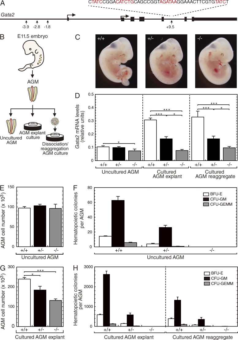
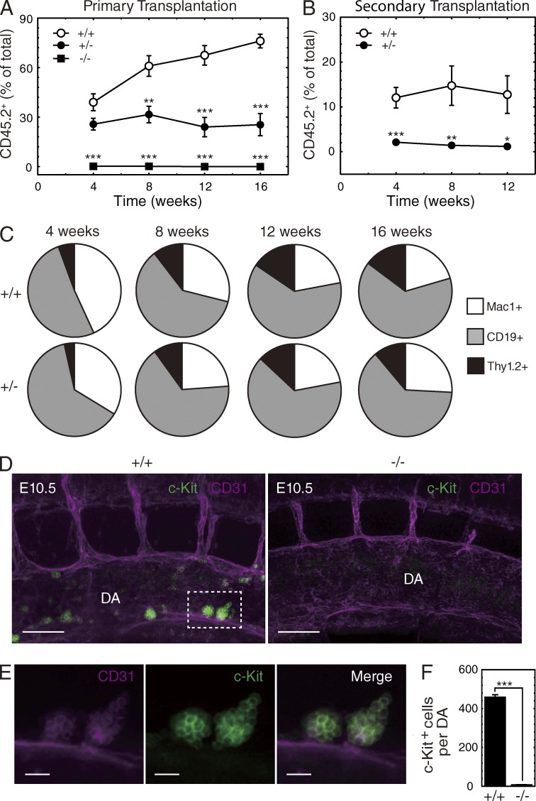
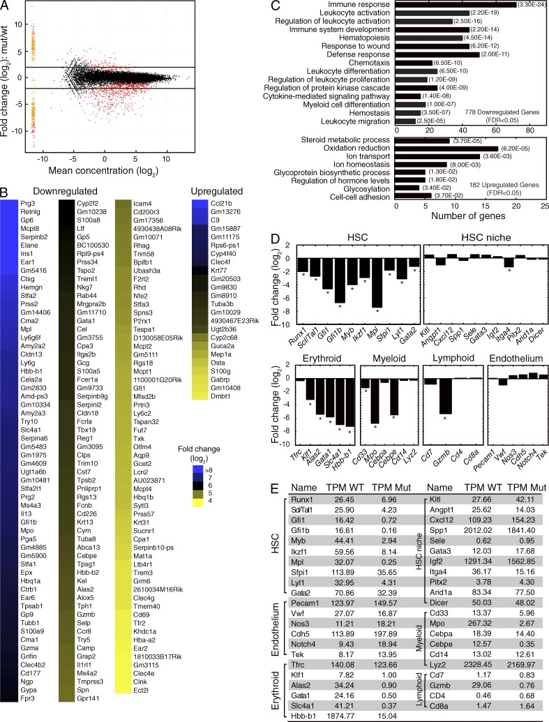
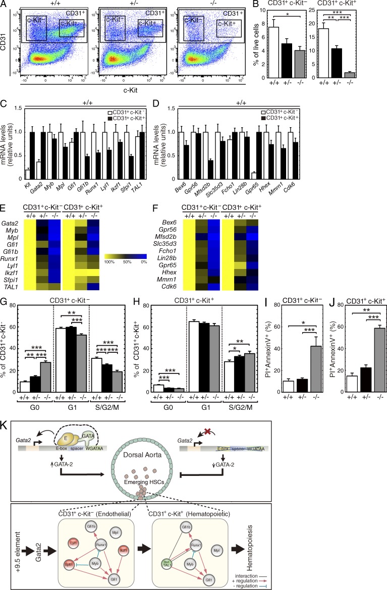
The generation of hematopoietic stem cells (HSCs) from hemogenic endothelium within the aorta, gonad, mesonephros (AGM) region of the mammalian embryo is crucial for development of the adult hematopoietic system. We described a deletion of a Gata2 cis-element (+9.5) that depletes fetal liver HSCs, is lethal at E13-14 of embryogenesis, and is mutated in an immunodeficiency that progresses to myelodysplasia/leukemia. Here, we demonstrate that the +9.5 element enhances Gata2 expression and is required to generate long-term repopulating HSCs in the AGM. Deletion of the +9.5 element abrogated the capacity of hemogenic endothelium to generate HSC-containing clusters in the aorta. Genomic analyses indicated that the +9.5 element regulated a rich ensemble of genes that control hemogenic endothelium and HSCs, as well as genes not implicated in hematopoiesis. These results reveal a mechanism that controls stem cell emergence from hemogenic endothelium to establish the adult hematopoietic system.


Figures

Figure 1.
Figure 1.
Cis-regulatory element requirement for Gata2 expression and hematopoietic progenitor activity in the AGM. (A) Schematic diagram for targeted deletion of the E-box-GATA composite element in murine Gata2 locus. (B) AGM ex vivo culture system. (C) Representative images of E11.5 +9.5+/+,+9.5+/−, and +9.5−/− embryos. (D) Real-time RT-PCR analysis of Gata2 expression levels in E11.5 uncultured AGMs, cultured AGM explants, and cultured AGM reaggregates (4 independent experiments and 5 litters for uncultured AGMs: +9.5+/+ [n = 13]; +9.5+/− [n = 18]; +9.5−/− [n = 7]; 2 independent experiments and 2 litters for cultured AGM explants: +9.5+/+ [n = 5]; +9.5+/− [n = 13]; +9.5−/− [n = 3]; 5 independent experiments and 5 litters for cultured AGM reaggregates: +9.5+/+ [n = 10]; +9.5+/− [n = 20]; +9.5−/− [n = 12]). (E and G) Quantitative analysis of cell number in E11.5 uncultured AGMs (E) and cultured AGM explants (G). (F and H) Quantitative analysis of colony-forming activity of hematopoietic progenitors in E11.5 uncultured AGMs (F), cultured AGM explants (H), and cultured AGM reaggregates (H; 2 independent experiments and 2 litters for uncultured AGMs: +9.5+/+ [n = 5]; +9.5+/− [n = 10]; +9.5−/− [n = 4]; 2 independent experiments and 2 litters for cultured intact AGM explants: +9.5+/+ [n = 5]; +9.5+/− [n = 5]; +9.5−/− [n = 3]; 2 independent experiments and 2 litters for cultured AGM reaggregates: +9.5+/+ [n = 6]; +9.5+/− [n = 8]; +9.5−/− [n = 6]). All error bars represent SEM. *, P < 0.05; ***, P < 0.001 (two-tailed unpaired Student’s t test).
Figure 2.
Figure 2.
Absence of long-term repopulating HSC activity and HSC generation in +9.5−/− AGM. (A) Contribution from +9.5+/− and +9.5−/− versus +9.5+/+ AGM explants (in a competitive transplantation assay +9.5+/+ [n = 11]; +9.5+/− [n = 11]; +9.5−/− [n = 6]). (B) Secondary transplant with bone marrow cells from primary recipient mice (+9.5+/+ [n = 8]; +9.5+/− [n = 8]). (C) The contribution of +9.5+/+ and +9.5+/− donor-derived CD45.2 cells to peripheral myeloid cells (Mac1+), B cells (CD19+) and T cells (Thy1.2+). (D) Whole-mount immunostaining of E10.5 +9.5+/+ (left) and +9.5−/− (right) embryos showing CD31+ cells (magenta) and c-Kit+ cells (green) within the aorta region. Bar, 100 µm. (E) Higher magnification of boxed region in D showing the representative aortic hematopoietic clusters in +9.5+/+ embryos. Bar, 20 µm. (F) Quantitation of c-Kit+ cells within the whole dorsal aorta (2 litters: +9.5+/+ [n = 3]; +9.5−/− [n = 3]). Error bars represent SEM. *, P < 0.05; **, P < 0.01; ***, P < 0.001 (two-tailed unpaired Student’s t test). Related to Videos 1 and 2.
Figure 3.
Figure 3.
A rich stem cell–regulatory genetic network controlled by a composite cis-element. RNA-seq-based comparison of gene expression in E11.5 +9.5+/+ (n = 3) and +9.5−/− (n = 3) cultured AGM explants. (A) MA plot for +9.5−/− versus +9.5+/+ cultured AGM explants. The red points indicate 778 and 182 genes down- or up-regulated (FDR < 0.05), respectively, in the mutants. (B) Heatmap depicting statistically significant, differentially expressed genes down- or up-regulated by >16 fold. (C) Gene Ontology analysis of genes down- or up-regulated in +9.5−/− cultured AGM explants with a cut-off FDR <0.05 using DAVID Bioinformatics Program (http://david.abcc.ncifcrf.gov/). The most enriched biological processes are shown with corresponding P values. (D) RNA-seq-based analysis of the expression levels of representative genes expressed in HSCs and the HSC niche that regulate hematopoiesis, as well as genes expressed in endothelium, erythroid, myeloid, and lymphoid cells (*, FDR < 0.05). (E) The relative expression levels (TPM, transcripts per million) of representative genes shown in the figure.
Figure 4.
Figure 4.
+9.5 element-regulated genetic network controls HSC emergence from hemogenic endothelium. (A) Representative flow cytometric plots depicting endothelial (CD31+c-Kit) and hematopoietic (CD31+c-Kit+) cells in cultured AGM explants. (B) Quantitative analysis of flow cytometry data expressed as percentage of live cells from cultured AGM explants (2 independent experiments and 4 litters: +9.5+/+ [n = 14]; +9.5+/− [n = 17]; +9.5−/− [n = 7]). (C and D) Quantitation of relative expression of HSC-related genes and representative novel genes in endothelial cells and hematopoietic cells from +9.5+/+ AGM explants. (E and F) Heatmap showing quantitation of relative expression of HSC-related genes and novel genes in endothelial cells and hematopoietic cells from +9.5+/+, +9.5+/−, and +9.5−/− AGM explants. (G and H) Cell cycle analysis of endothelial (CD31+c-Kit) and hematopoietic cells (CD31+c-Kit+) from +9.5+/+, +9.5+/−, and +9.5−/− AGM explants (thr3e independent experiments and 5 litters: +9.5+/+ [n = 10]; +9.5+/− [n = 22]; +9.5−/− [n = 11]). (I and J) Flow cytometric analysis of the percentage of apoptotic cells (PI+Annexin V+) within the endothelial cells (CD31+c-Kit) and hematopoietic cells (CD31+c-Kit+) from +9.5+/+, +9.5+/−, and +9.5−/− AGM explants (2 independent experiments and 2 litters: +9.5+/+ [n = 3]; +9.5+/− [n = 10]; +9.5−/− [n = 4]). (K) Model describing +9.5 cis-element requirement for HSC emergence from AGM and +9.5 cis-element–dependent genetic networks in AGM endothelial and hematopoietic cells. Error bars represent SEM. *, P < 0.05; **, P < 0.01; ***, P < 0.001 (two-tailed unpaired Student’s t test).

References

    1. Bertrand J.Y., Chi N.C., Santoso B., Teng S., Stainier D.Y., Traver D. 2010. Haematopoietic stem cells derive directly from aortic endothelium during development. Nature. 464:108–111 10.1038/nature08738 - DOI - PMC - PubMed
    1. Boisset J.C., van Cappellen W., Andrieu-Soler C., Galjart N., Dzierzak E., Robin C. 2010. In vivo imaging of haematopoietic cells emerging from the mouse aortic endothelium. Nature. 464:116–120 10.1038/nature08764 - DOI - PubMed
    1. Bresnick E.H., Lee H.Y., Fujiwara T., Johnson K.D., Keles S. 2010. GATA switches as developmental drivers. J. Biol. Chem. 285:31087–31093 10.1074/jbc.R110.159079 - DOI - PMC - PubMed
    1. Bresnick E.H., Katsumura K.R., Lee H.Y., Johnson K.D., Perkins A.S. 2012. Master regulatory GATA transcription factors: mechanistic principles and emerging links to hematologic malignancies. Nucleic Acids Res. 40:5819–5831 10.1093/nar/gks281 - DOI - PMC - PubMed
    1. Chen M.J., Yokomizo T., Zeigler B.M., Dzierzak E., Speck N.A. 2009. Runx1 is required for the endothelial to haematopoietic cell transition but not thereafter. Nature. 457:887–891 10.1038/nature07619 - DOI - PMC - PubMed

Publication types

Substances