. 2013 Dec 16;210(13):2843-50.
doi: 10.1084/jem.20130751. Epub 2013 Dec 2.

Gata2 is required for HSC generation and survival

Affiliations

Gata2 is required for HSC generation and survival

Emma de Pater et al. J Exp Med. .

Abstract

Knowledge of the key transcription factors that drive hematopoietic stem cell (HSC) generation is of particular importance for current hematopoietic regenerative approaches and reprogramming strategies. Whereas GATA2 has long been implicated as a hematopoietic transcription factor and its dysregulated expression is associated with human immunodeficiency syndromes and vascular integrity, it is as yet unknown how GATA2 functions in the generation of HSCs. HSCs are generated from endothelial cells of the major embryonic vasculature (aorta, vitelline, and umbilical arteries) and are found in intra-aortic hematopoietic clusters. In this study, we find that GATA2 function is essential for the generation of HSCs during the stage of endothelial-to-hematopoietic cell transition. Specific deletion of Gata2 in Vec (Vascular Endothelial Cadherin)-expressing endothelial cells results in a deficiency of long-term repopulating HSCs and intra-aortic cluster cells. By specific deletion of Gata2 in Vav-expressing hematopoietic cells (after HSC generation), we further show that GATA2 is essential for HSC survival. This is in contrast to the known activity of the RUNX1 transcription factor, which functions only in the generation of HSCs, and highlights the unique requirement for GATA2 function in HSCs throughout all developmental stages.


Figures
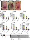
Figure 1.
Figure 1.
GATA2 is required in VE-Cadherin–expressing cells for hematopoietic progenitor formation. (A) Bright field images of E14 WT, Vec-Cre:Gata2f/f, and Vav-Cre:Gata2f/f embryos. Bar, 2 mm. (B) CFU-C numbers per AGM and FL from Gata2-deleted embryos. Germline Gata2−/− AGMs at E10 (30–34 sp), n = 4. The number of embryos analyzed is 8 WT, 11 Gata2+/−, and 4 Gata2−/−. For Vec-Cre–mediated Gata2 deletion: WT, Vec-Cre:Gata2f/+, and Vec-Cre:Gata2f/f AGMs and FLs at E10 (30–36 sp), E11 (43–48 sp), and FL E14, n = 2. The number of E10 embryos analyzed is 5 WT, 6 Vec-Cre:Gata2f/+, and 6 Vec-Cre:Gata2f/f ; n = 3. The number of E11 embryos analyzed is 6 WT, 3 Vec-Cre:Gata2f/+, and 4 Vec-Cre:Gata2f/f; E14 FL n = 3. The number of embryos analyzed is 11 WT, 3 Vec-Cre:Gata2f/+, and 5 Vec-Cre:Gata2f/f. Error bars are mean ± SEM of total colony numbers. *, P < 0.05; **, P < 0.01, significance determined by Student’s t test. ee, embryo equivalent of specified tissue. (C) Schematic representation of the Gata2 locus with Loxp sites and genotyping PCR primer location. Loxp sites are indicated by orange triangles. Gel showing the Gata2 flox band of 844 bp (f) deleted (Δ) band of 181 bp and WT Gata2 band (+) of 717 bp for individual CFU-C. The number of identified genotypes of individual colonies is shown beneath the gel.
Figure 2.
Figure 2.
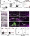
GATA2 is required in VE-Cadherin–expressing cells for the generation of HSCs and vascular hematopoietic cluster cells. (A) Graph showing the percentage of peripheral blood donor cell chimerism in adult recipients at 4 mo after transplantation. Recipients were injected with AGM cells from WT (1–2 ee; 3 of 10 recipients repopulated, n = 3), Vec-Cre:Gata2f/+ (1–2 ee; 0 of 7 recipients repopulated), or Vec-Cre:Gata2f/f (1–3 ee; 0 of 7 recipients repopulated) E11 embryos. Reconstitution kinetics showed similar outcomes at 1, 2, 3, and 4 mo after transplantation. (B) FACS analysis of LinCD48 gated, cKit- and Sca1-stained E14 FL cells from WT and Vec-Cre:Gata2f/f embryos. Percentage of cKit+Sca1+ cells within the LinCD48 population is shown for WT (0.019 ± 0.01%) and Vec-Cre:Gata2f/f (0.16 ± 0.04%, P < 0.05). The right panel shows FACS analysis of LSKCD48 gated, CD150- and Annexin V–stained Vec-Cre:Gata2f/f E14 FLs. Phenotypic HSCs are indicated within the red quadrants. 100% of LSK SLAM cells are Annexin V–positive (n = 2, number of embryos analyzed = 7 WT, 5 Vec-Cre:Gata2f/f; significance determined by Student’s t test). (For WT, see Fig. 3 E.) (C) Immunostaining of E10 (30/31 sp) embryos showing cKit+ hematopoietic cluster cells in germline Gata2+/+, Gata2+/−, and Gata2−/− aortae. Arrowheads indicate some of the hematopoietic cluster cells along the aortic wall. Bar, 50 µm. DA = dorsal aorta, PGC = primordial germ cells (n = 1). (D) Whole mount immunostaining of E10.5 (36/37 sp) Gata2f/+ and Vec-Cre:Gata2f/f embryos showing CD31+ vascular endothelial cells (magenta) and cKit+ hematopoietic cluster cells (green). The top panels show the dorsal aorta (DA), middle panels the umbilical artery (U), and bottom panels a high-magnification image of a cKit+ cluster and aortic endothelium. Arrowheads indicate a few cKit+ hematopoietic cluster cells and arrow indicates a flat cKit+ cell embedded in the endothelium (n = 2, number of embryos analyzed is 2 WT and 2 Vec-Cre:Gata2f/f; significance determined by Student’s t test). Bars: (top and middle) 100 µm; (bottom) 10 µm. (E) FACS analysis showing Flk1- and cKit-stained WT and Vec-Cre:Gata2f/f E11 AGMs. Flk1+ cKit+ cells (red quadrant) and gating strategy for the viability (right) are shown. HoechstAnnexin V cells are viable, HoechstAnnexin V+ are early apoptotic cells, Hoechst+Annexin V+ are late apoptotic cells, and Hoechst+Annexin V cells are dead (n = 3; number of embryos analyzed = 7 WT and 6 Vec-Cre:Gata2f/f; significance determined by Student’s t test).
Figure 3.
Figure 3.
Gata2 is required in HPCs and in HSCs for survival after their generation. (A) Graphs showing the CFU-C numbers per WT, Vav-Cre:Gata2f/+, or Vav-Cre:Gata2f/f embryo equivalent (ee) of AGM and FL cells. Error bars indicate mean ± SEM. *, P < 0.05; **, P < 0.01. The number of E11 AGMs and FLs analyzed is 11 WT, 12 f/+, and 6 f/f; n = 4. For E11 AGM, P < 0.05, and E11 FL, P < 0.01. For E14 FL (P < 0.01; n = 3), the number of embryos analyzed = 10 WT, 5 f/+, and 20 f/f. Significance was determined by Student’s t test. (B) FACS analysis of WT, Vav-Cre:Gata2f/+, and Vav-Cre:Gata2f/f E14 FL for LSK SLAM markers. LinCD48CD150+ gated cells were analyzed for cKit and Sca1. Percentages of LSK SLAM cells (gated region) are shown. (C) Graph of the percentage of total CFU-C and CFU-GEMM per 100 LSK SLAM cells of each genotype (n = 3). The number of embryos analyzed = 3 WT, 3 f/+, and 3 f/f. Error bars are mean ± SEM of total colony numbers and of CFU-GEMM only. *, P < 0.05; **, P < 0.01; significance determined by Student’s t test. (D) Graph showing the percentage of peripheral blood donor cell chimerism in adult recipients injected with 105 WT (n = 2; 9 of 9 recipients repopulated) or 105 Vav-Cre:Gata2f/f E14 FL cells (0 of 7 recipients repopulated, P < 0.01). Diamonds represent individual recipients, with the mean represented by a horizontal bar. Significance was determined by Student’s t test. (E) Annexin V FACS analysis of WT and Vav-Cre:Gata2f/f E16 FL LSK SLAM cells. LSKCD48 gated cells were analyzed for Annexin V and CD150 (n = 2). The number of embryos analyzed = 3 WT and 6 f/f. Phenotypic HSCs (red quadrant) and the percentages of apoptotic and viable cells are indicated. (Not depicted: for E14 FL n = 3; the number of embryos analyzed = 8 WT, 10 f/+, and 9 f/f.)

References

    1. Boisset J.C., van Cappellen W., Andrieu-Soler C., Galjart N., Dzierzak E., Robin C. 2010. In vivo imaging of haematopoietic cells emerging from the mouse aortic endothelium. Nature. 464:116–120 10.1038/nature08764 - DOI - PubMed
    1. Boisset J.C., Clapes T., Van Der Linden R., Dzierzak E., Robin C. 2013. Integrin αIIb (CD41) plays a role in the maintenance of hematopoietic stem cell activity in the mouse embryonic aorta. Biol. Open. 2:525–532 10.1242/bio.20133715 - DOI - PMC - PubMed
    1. Charles M.A., Saunders T.L., Wood W.M., Owens K., Parlow A.F., Camper S.A., Ridgway E.C., Gordon D.F. 2006. Pituitary-specific Gata2 knockout: effects on gonadotrope and thyrotrope function. Mol. Endocrinol. 20:1366–1377 10.1210/me.2005-0378 - DOI - PubMed
    1. Chen M.J., Yokomizo T., Zeigler B.M., Dzierzak E., Speck N.A. 2009. Runx1 is required for the endothelial to haematopoietic cell transition but not thereafter. Nature. 457:887–891 10.1038/nature07619 - DOI - PMC - PubMed
    1. de Bruijn M.F., Speck N.A., Peeters M.C., Dzierzak E. 2000. Definitive hematopoietic stem cells first develop within the major arterial regions of the mouse embryo. EMBO J. 19:2465–2474 10.1093/emboj/19.11.2465 - DOI - PMC - PubMed

Publication types

Substances