Review
. 2013 Dec 1;27(23):2513-30.
doi: 10.1101/gad.229559.113.

Chromothripsis and beyond: rapid genome evolution from complex chromosomal rearrangements

Affiliations
Review

Chromothripsis and beyond: rapid genome evolution from complex chromosomal rearrangements

Cheng-Zhong Zhang et al. Genes Dev. .

Abstract

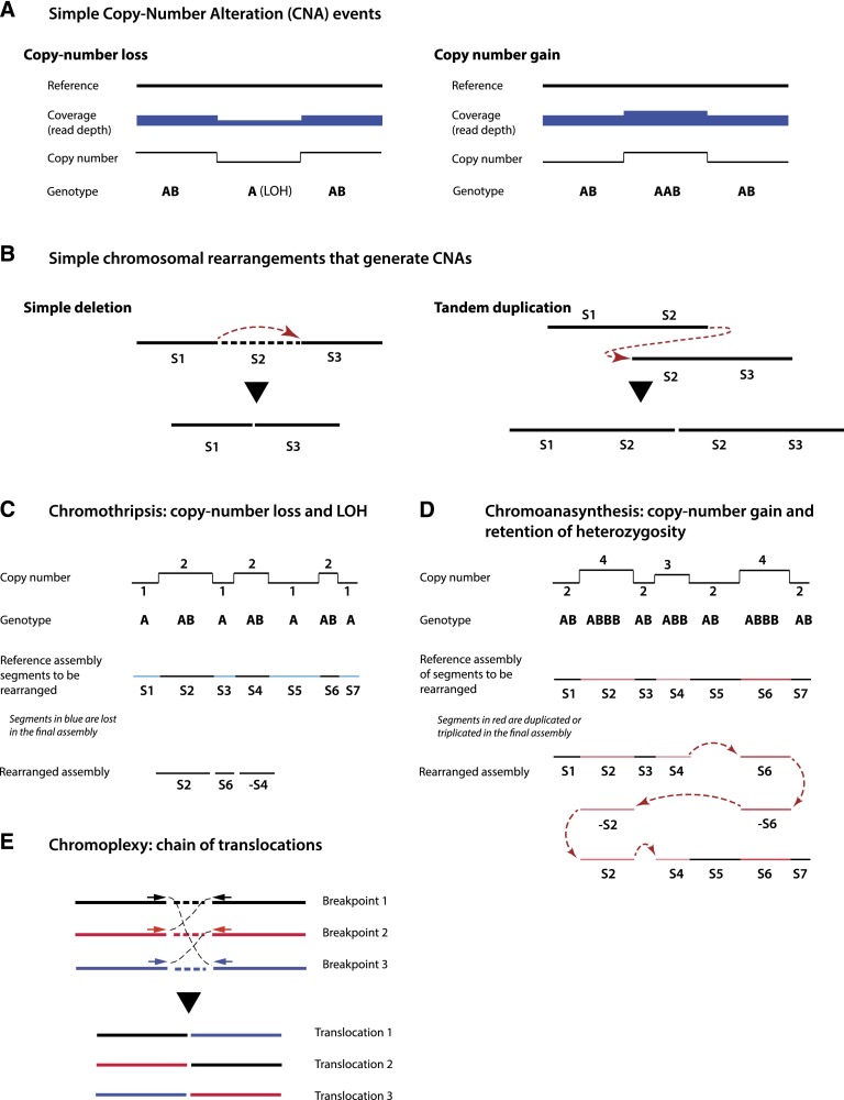
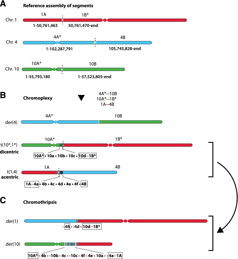
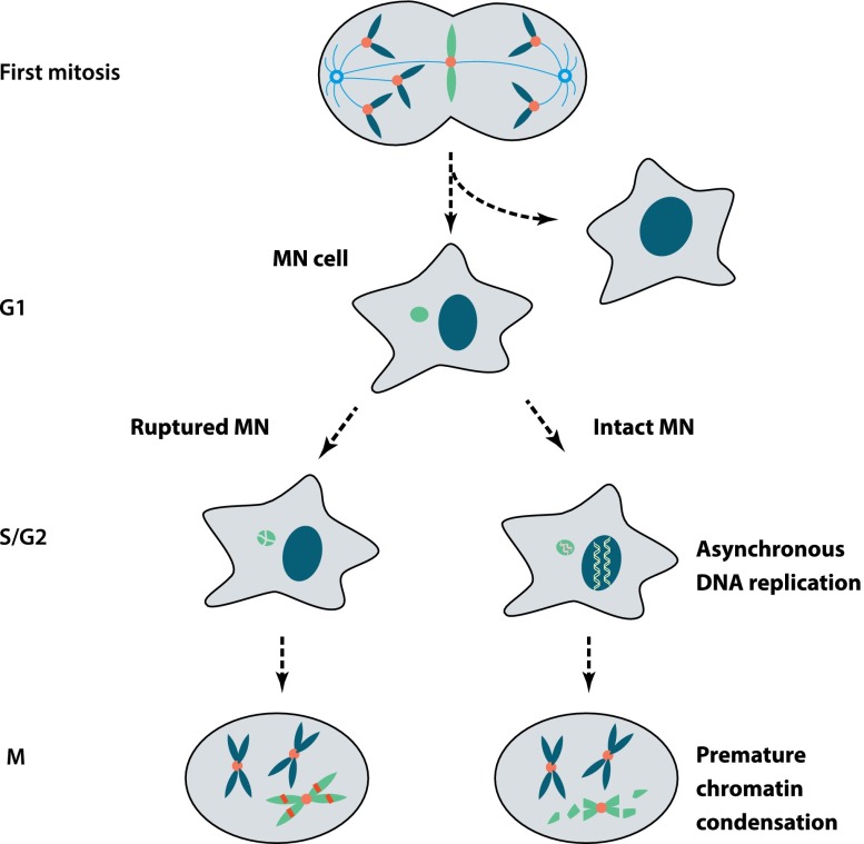
Recent genome sequencing studies have identified several classes of complex genomic rearrangements that appear to be derived from a single catastrophic event. These discoveries identify ways that genomes can be altered in single large jumps rather than by many incremental steps. Here we compare and contrast these phenomena and examine the evidence that they arise "all at once." We consider the impact of massive chromosomal change for the development of diseases such as cancer and for evolution more generally. Finally, we summarize current models for underlying mechanisms and discuss strategies for testing these models.

Keywords: cancer; chromoanasynthesis; chromoplexy; chromosomal translocation; chromothripsis; copy number alteration; genome evolution.


Figures

Figure 1.
Figure 1.
Characteristics of simple and complex chromosomal rearrangements. (A) Copy number losses or gains can be detected by array-based or sequencing-based technologies from abrupt changes in the read depth signal (sequencing) or the intensity of fluorescent probes (array). Loss or gain generally affects only one homolog; therefore, a copy number loss also results in LOH. (B) Copy number alterations are often generated by simple chromosomal rearrangements, such as deletions or tandem duplications. (C) Chromothripsis is characterized by an alternating copy number profile with loss and retention of heterozygosity. Minus signs in C and D denote chromosomal segments that are inverted. Fluorescent in situ hybridization (FISH) characterization confirms that only one chromatid (allele B) is affected. (D) Chromoanasynthesis is characterized by a copy number profile that alternates between euploid and higher ploidy. In contrast to chromothripsis, chromoanasynthesis does not necessarily exhibit LOH but reflects resynthesis of segments from one chromatid. Chromoanasynthesis also contains frequent insertions of short sequences between the rearrangement junctions (red arrows) that are copied from the rearranged segments (templated insertions). Both DNA resynthesis and templated insertions are suggestive of replication-based mechanisms. (E) Chromoplexy is characterized by a closed chain of translocations, with little or no copy number alteration.
Figure 2.
Figure 2.
Signatures of chromosomal rearrangements from paired-end sequencing. (A) Four major types of chromosomal rearrangement signatures deduced from paired-end sequencing: deletion type, tandem duplication type, inversion type, and long-range type (including interchromosomal). Each type of read pair shows aberrations from proper pairs when aligned to the reference assembly, which implies a new junction in the test chromosome. A deletion (read pairs shown as red arrows) or tandem duplication (shown as green arrows) only introduce one fusion and hence can be unambiguously inferred from a single type of signature (in the absence of other overlapping events). Inversion events, however, introduce at least two rearranged junctions (shown here is a simple inversion); hence, the rearranged genome can only be constructed by considering all related events. The same is true for long-range events with pair mates residing in different chromosomes or separated by centromeres, assembly gaps, or other rearrangements. In general, it is necessary to incorporate copy number information together with rearrangement signatures to derive the structure of the rearranged genome (for a computational framework, see Greenman et al. 2012a). (B) Examples of complex chromosomal rearrangements exhibiting multiple overlapping rearrangement signatures that do not reflect the same events as the “typing.” The discordant read pair signatures (color arrows) are shown both in the rearranged genome and according to their alignment to the reference genome. (i) A complex rearrangement resulting in the deletion of segment A and swapping of segments B and C generates two “tandem duplication” and one “deletion” read pair signatures, none of which corresponds to a simple event. (ii) Two inversions alter a deletion signature into a tandem duplication signature (green arrows).
Figure 3.
Figure 3.
Reconstruction of a local complex rearrangement and its mutational history from read pair signatures and copy number information. (A) Copy number alterations can be detected by array methods or read depths in sequencing data; rearrangement signatures can be detected from discordant read pairs in sequencing data. (B) By combining copy number information and rearrangement signatures, one can construct the configuration of the rearranged segments in the chromothriptic chromosome. (C) The history of mutation events that could have generated the outcome in B is often not unique. However, all solutions have to break four junctions in the reference assembly (L|A, A|B, B|C, and C|R) and generate three new junctions (L|C, C|B, and B|R). A catastrophe model can easily solve this puzzle because all of the breaks and translocations occur all at once. By comparison, a solution from the gradual model of simple events requires at least three simple events to generate the final configuration (one for each new junction). As each event causes two breakpoints, three events need six breakpoints, which requires two additional breaks in addition to the four original junctions to be broken. These two additional breaks arise from either within the lost A segment (lost history) or hitting the same junction multiple times (recurrent breaks). Shown here is one possible solution that involves recurrent breaks at both B|C and C|R junctions. Both lost history and recurrent breaks are highly unlikely. As the number of rearranged fragments increases, the need to include these types of unlikely events further increases, strongly arguing against the gradual model.
Figure 4.
Figure 4.
Chromoplexy and ChainFinder. (A) Chromoplexy is an extension of balanced rearrangements to multiple partners. The different colors of linked discordant pair mates reflect different chromosomes or loci to which each mate is aligned. DNA resection during the repair of DSBs often results in loss of a small segment between the two ends. (B) Adjacent translocations of two breakpoint ends can arise in two different ways. (Left) In a single-event model, a single DNA DSB generates two broken ends with a small deletion due to DNA resection; joining of the two broken ends with ends from other DNA breaks produces the observed translocations. (Right) In a multiple-event model, two independent balanced translocations occur independently at different times; the inner DNA segment could have been lost in a subsequent deletion event (shown in the dashed box) or due to DNA resection. The distance between the two adjacent translocation ends, D, provides a statistical measure of the likelihood of either scenario. The two-event model implies that two independent breaks must have occurred within distance D. This likelihood is approximately given by 1 − exp(−μD), where μ is the average density of chromosomal breaks that can be estimated for each sample at any given locus (≲10−6 per base pair). When the ends are very close (100–1000 bp), this probability is vanishingly small, favoring the single-event model and rejecting the multievent model. The central idea of ChainFinder is to find “chained” translocations that form a closed cycle and where the multiple-event model can be rejected for any two pairs of adjacent translocations (i.e., no reciprocal translocation can be postulated as an intermediate state, connecting two smaller chains).
Figure 5.
Figure 5.
Possible decomposition of rearrangements reported by Kloosterman et al. (2011a) into two chains. The first chain is plausibly a chromoplexy event that swaps partners between six chromosomal arms, shown in A. Asterisks denote chromosomal segments containing a centromere. Newly formed junctions are shown as red links. This chromoplexy episode would create one functional (single centromere-containing) derivative chromosome, t(4p*, 10q), plus two aberrant ones, t(10p*,1q*) (dicentric) and t(1p,4q) (acentric), as shown in B. The unstable dicentric and acentric chromosomes could then have triggered the remaining rearrangements shown in C, which are interlinked and resemble a chromothripsis. The outcome of this second chain is the swap of partners between the two nonfunctional chromosomes 10A*–1B* and 1A–4B, which results in two new derivative chromosomes, 10A*–(-1A) and (-4B)–1B*. This event is accompanied by fragmentation and repair of multiple segments near the junctions from the first chromoplexy event (10a, 10b, and 10c; and 4b, 4c, 4d, 4e, and 4f).
Figure 6.
Figure 6.
Extensive DNA damage of chromosomes in micronuclei (MN). Micronuclei can form by missegregation of whole chromosomes after mitotic errors. Pictured here is a “merotelic” kinetochore attachment where a single intact chromatid is caught in a tug-of-war between microtubules from opposite poles. Micronuclei can also originate from chromosome breaks, generating acentric chromosome fragments, or be generated from chromosome bridges by a mechanism that has not been determined (Hoffelder et al. 2004). (Left) Newly generated micronuclei can undergo irreversible loss of nuclear membrane integrity (Hatch et al. 2013). If this occurs during S phase, chromosomes in micronuclei will acquire massive DNA damage. (Right) Alternatively, late-replicating chromosomes in micronuclei may undergo DNA damage if the cell enters mitosis before micronuclear replication is complete (Crasta et al. 2012), subjecting the chromosome to premature chromosome compaction (PCC). The extent and mechanism of DNA damage during PCC remains to be established. During the subsequent cell division cycle, the micronuclear genome can be reincorporated into the primary nucleus, remain isolated in a micronucleus, or possibly be lost due to autophagy or extrusion.

Similar articles

Cited by

  • The tandem duplicator phenotype as a distinct genomic configuration in cancer.
    Menghi F, Inaki K, Woo X, Kumar PA, Grzeda KR, Malhotra A, Yadav V, Kim H, Marquez EJ, Ucar D, Shreckengast PT, Wagner JP, MacIntyre G, Murthy Karuturi KR, Scully R, Keck J, Chuang JH, Liu ET. Menghi F, et al. Proc Natl Acad Sci U S A. 2016 Apr 26;113(17):E2373-82. doi: 10.1073/pnas.1520010113. Epub 2016 Apr 7. Proc Natl Acad Sci U S A. 2016. PMID: 27071093 Free PMC article.
  • FANCA safeguards interphase and mitosis during hematopoiesis in vivo.
    Abdul-Sater Z, Cerabona D, Potchanant ES, Sun Z, Enzor R, He Y, Robertson K, Goebel WS, Nalepa G. Abdul-Sater Z, et al. Exp Hematol. 2015 Dec;43(12):1031-1046.e12. doi: 10.1016/j.exphem.2015.08.013. Epub 2015 Sep 11. Exp Hematol. 2015. PMID: 26366677 Free PMC article.
  • An Organismal CNV Mutator Phenotype Restricted to Early Human Development.
    Liu P, Yuan B, Carvalho CMB, Wuster A, Walter K, Zhang L, Gambin T, Chong Z, Campbell IM, Coban Akdemir Z, Gelowani V, Writzl K, Bacino CA, Lindsay SJ, Withers M, Gonzaga-Jauregui C, Wiszniewska J, Scull J, Stankiewicz P, Jhangiani SN, Muzny DM, Zhang F, Chen K, Gibbs RA, Rautenstrauss B, Cheung SW, Smith J, Breman A, Shaw CA, Patel A, Hurles ME, Lupski JR. Liu P, et al. Cell. 2017 Feb 23;168(5):830-842.e7. doi: 10.1016/j.cell.2017.01.037. Cell. 2017. PMID: 28235197 Free PMC article.
  • Endogenous DNA Damage as a Source of Genomic Instability in Cancer.
    Tubbs A, Nussenzweig A. Tubbs A, et al. Cell. 2017 Feb 9;168(4):644-656. doi: 10.1016/j.cell.2017.01.002. Cell. 2017. PMID: 28187286 Free PMC article. Review.
  • Single-cell analysis of structural variations and complex rearrangements with tri-channel processing.
    Sanders AD, Meiers S, Ghareghani M, Porubsky D, Jeong H, van Vliet MACC, Rausch T, Richter-Pechańska P, Kunz JB, Jenni S, Bolognini D, Longo GMC, Raeder B, Kinanen V, Zimmermann J, Benes V, Schrappe M, Mardin BR, Kulozik AE, Bornhauser B, Bourquin JP, Marschall T, Korbel JO. Sanders AD, et al. Nat Biotechnol. 2020 Mar;38(3):343-354. doi: 10.1038/s41587-019-0366-x. Epub 2019 Dec 23. Nat Biotechnol. 2020. PMID: 31873213 Free PMC article.

References

    1. Adey A, Burton JN, Kitzman JO, Hiatt JB, Lewis AP, Martin BK, Qiu R, Lee C, Shendure J 2013. The haplotype-resolved genome and epigenome of the aneuploidy HeLa cancer cell line. Nature 500: 207–211 - PMC - PubMed
    1. Alexandrov LB, Nik-Zainal S, Wedge DC, Aparicio SA, Behjati S, Biankin AV, Bignell GR, Bolli N, Borg A, Borrensen-Dale AL, et al. 2013. Signatures of mutational processes in human cancer. Nature 500: 415–421 - PMC - PubMed
    1. Alt FW, Zhang Y, Meng FL, Guo C, Schwer B 2013. Mechanisms of programmed DNA lesions and genomic instability in the immune system. Cell 152: 417–429 - PMC - PubMed
    1. Anand RP, Lovett ST, Haber JE 2013. Break-induced DNA replication. Cold Spring Harb Perspect Biol doi: 10.1101/cshperspect.a010397 - PMC - PubMed
    1. Arlt MF, Mulle JG, Schaibley VM, Ragland RL, Durkin SG, Warren ST, Glover TW 2009. Replication stress induces genome-wide copy number changes in human cells that resemble polymorphic and pathogenic variants. Am J Hum Genet 84: 339–350 - PMC - PubMed

Publication types