. 2013 Dec 1;27(23):2537-42.
doi: 10.1101/gad.226373.113.

Deregulated origin licensing leads to chromosomal breaks by rereplication of a gapped DNA template

Affiliations

Deregulated origin licensing leads to chromosomal breaks by rereplication of a gapped DNA template

Kai J Neelsen et al. Genes Dev. .

Abstract

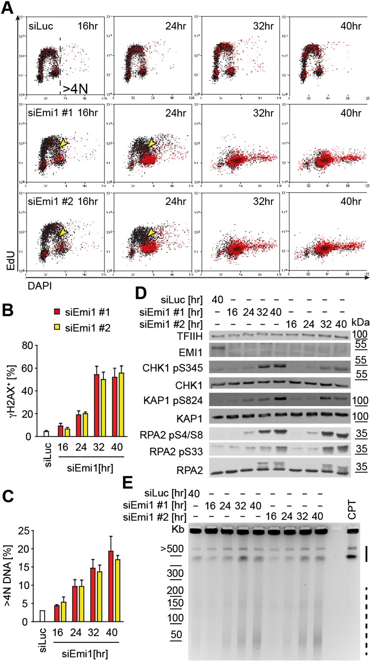
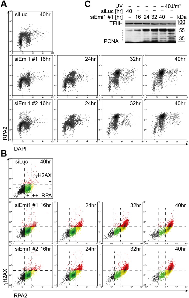
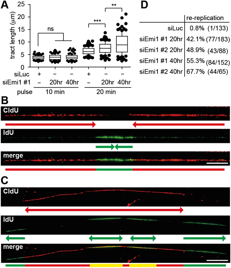
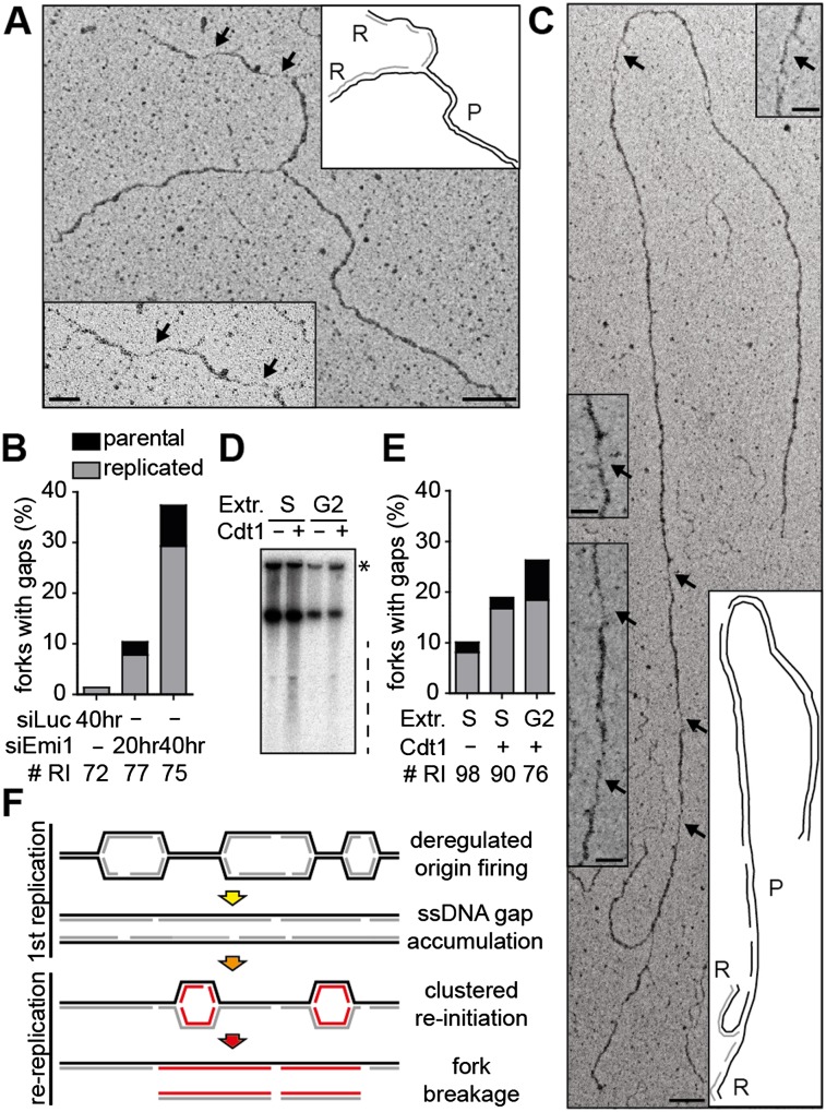
Deregulated origin licensing and rereplication promote genome instability and tumorigenesis by largely elusive mechanisms. Investigating the consequences of Early mitotic inhibitor 1 (Emi1) depletion in human cells, previously associated with rereplication, we show by DNA fiber labeling that origin reactivation occurs rapidly, well before accumulation of cells with >4N DNA, and is associated with checkpoint-blind ssDNA gaps and replication fork reversal. Massive RPA chromatin loading, formation of small chromosomal fragments, and checkpoint activation occur only later, once cells complete bulk DNA replication. We propose that deregulated origin firing leads to undetected discontinuities on newly replicated DNA, which ultimately cause breakage of rereplicating forks.

Keywords: DNA replication; genome integrity; origin licensing; rereplication; tumorigenesis.


Figures

Figure 1.
Figure 1.
Emi1 depletion causes DNA replication stress in S phase and DDR activation and DNA breakage in cells with ≥4 DNA. (A) FACS analysis of DNA synthesis (EdU), DNA content (DAPI), and DDR activation (γH2AX) after mock (siLUC) or Emi1 depletion in U2OS cells using two different siRNAs. γH2AX+ cells are in red (see also Supplemental Fig. S1A). Yellow arrowheads indicate cells with compromised DNA synthesis. γH2AX+ cells (B) and cells with >4N DNA (C) after mock (siLUC) or Emi1 depletion quantified by FACS. Mean + SEM; n = 3. (D) ATR (pCHK1), ATM (pKAP1) activation, RPA phosphorylation (RPA2 pS4/S8 and pS33), and total DDR proteins (CHK1, KAP1, and RPA2) assessed by Western blot upon Emi1 depletion. (TFIIH) Loading control. (E) DNA breakage after mock (siLUC) or Emi1 depletion monitored by pulse-field gel electrophoresis. The solid and dashed lines indicate large (0.5- to 2-Mb) and smaller (20- to 100-kb) chromosomal fragments, respectively. The molecular size markers are based on data in Supplemental Figure S1B. Four-hour treatment with 1 μM camptothecin (CPT) served as a positive control for DSB.
Figure 2.
Figure 2.
siEmi1-induced deregulation of origin licensing promotes RPA chromatin binding and ubiquitylation of PCNA from the first S phase. (A) FACS analysis of chromatin-bound RPA and DNA content (DAPI) after mock (siLuc) or Emi1 depletion in U2OS cells. (B) γH2AX/RPA levels in samples in A. Black, green, and yellow regions identify RPA negative cells (−), cells with S-phase RPA levels (+), and cells with elevated RPA (++), respectively. The red region identifies γH2AX+ cells. See Supplemental Figure S4B for Emi1 levels. (C) Analysis of PCNA ubiquitylation in mock-transfected cells (siLuc) and at the indicated time points after Emi1 depletion (siEmi1 #1). The dotted line indicates ubiquitylated PCNA. UV-irradiated cells served as positive control. (TFIIH) Loading control.
Figure 3.
Figure 3.
Rereplication is detectable by a DNA fiber-spreading assay before completion of bulk DNA synthesis. (A) Length of newly replicated tracts (IdU; green) in mock-depleted U2OS cells (siLuc) and after Emi1 depletion (siEmi1 #1), using 10-min or 20-min labeling pulses. (B,C) Representative DNA tracts labeled with CldU for 2 h and IdU for 30 min to identify termination and rereplication events. (B) A replication “termination” event. (C) Two “rereplication” events in close proximity. (D) Quantification of rereplication/termination events as shown in B and C after mock (siLuc) or Emi1 depletion. The percentage indicated represents the fraction of rereplication events in the total population of “red–green–red” tracts analyzed. (Whiskers) 10–90 percentile; (***) P < 0.0001; (**) P < 0.005; (ns) not significant, Mann-Whitney test; n = 100 in A. Bar, 10 μm. See Supplemental Figure S5, A and B, for Emi1 levels and labeling protocols to study fork progression (A) and rereplication events (B,C).
Figure 4.
Figure 4.
Emi1 depletion leads to ssDNA gaps on the replicated duplex, which persist as a template for rereplicating forks. (A,C) Electron micrographs of representative replication forks from U2OS cells 40 h after transfection with siEmi1. Black arrows indicate ssDNA gaps. The insets show magnified ssDNA gaps and schemes of fork structure, indicating parental (P) and replicated (R) duplexes. Gaps are on a replicated duplex in A and on the parental duplex in C. Black and gray lines describe parental and newly synthesized DNA strands in the replicated duplexes, respectively. Bars: 100 nm (250 base pairs [bp]); inset, 50 nm. (B) Frequency of replication forks with ssDNA gaps in mock-depleted cells (siLuc) and after Emi1 depletion (siEmi1 #1). #RI is the number of analyzed replication intermediates. (D) Sperm nuclei replication assays in Xenopus interphase extracts. For S-phase experiments, extracts were optionally supplemented with 10 ng/μL Cdt1 at the time of sperm and [α-32P]dATP addition and incubated for 60 min. For G2 experiments, Cdt1 was optionally added with [α-32P]dATP 90 min after sperm addition and incubated for a further 60 min. After incubation, DNA was isolated, separated by neutral agarose gel electrophoresis, and autoradiographed. The dashed line indicates sperm DNA fragmentation. The asterisk indicates branched replicating DNA molecules retained in the well. (E) Frequency of replication forks with ssDNA gaps recovered after sperm nuclei incubation in S-phase or G2-phase extracts (see D), with optional addition of Cdt1. # RI is the number of analyzed replication intermediates. (F) Model for the formation of chromosomal breaks upon deregulation of origin licensing by Emi1 depletion. Excessive firing of clustered origins leads to replication stress during the first S phase and accumulation of ssDNA gaps. Uncontrolled reactivation of replication origins in this context triggers chromosomal breakage by replication of a discontinuous template.

References

    1. Arias EE, Walter JC 2006. PCNA functions as a molecular platform to trigger Cdt1 destruction and prevent re-replication. Nat Cell Biol 8: 84–90 - PubMed
    1. Arias EE, Walter JC 2007. Strength in numbers: Preventing rereplication via multiple mechanisms in eukaryotic cells. Genes Dev 21: 497–518 - PubMed
    1. Bester AC, Roniger M, Oren YS, Im MM, Sarni D, Chaoat M, Bensimon A, Zamir G, Shewach DS, Kerem B 2011. Nucleotide deficiency promotes genomic instability in early stages of cancer development. Cell 145: 435–446 - PMC - PubMed
    1. Blow JJ, Dutta A 2005. Preventing re-replication of chromosomal DNA. Nat Rev Mol Cell Biol 6: 476–486 - PMC - PubMed
    1. Blow JJ, Gillespie PJ 2008. Replication licensing and cancer—a fatal entanglement? Nat Rev Cancer 8: 799–806 - PMC - PubMed

Publication types

LinkOut - more resources