. 2013 Dec 2;369(1633):20130144.
doi: 10.1098/rstb.2013.0144. Print 2014 Jan 5.

Microtubule-associated protein tau is essential for long-term depression in the hippocampus

Affiliations

Microtubule-associated protein tau is essential for long-term depression in the hippocampus

Tetsuya Kimura et al. Philos Trans R Soc Lond B Biol Sci. .

Abstract

The microtubule-associated protein tau is a principal component of neurofibrillary tangles, and has been identified as a key molecule in Alzheimer's disease and other tauopathies. However, it is unknown how a protein that is primarily located in axons is involved in a disease that is believed to have a synaptic origin. To investigate a possible synaptic function of tau, we studied synaptic plasticity in the hippocampus and found a selective deficit in long-term depression (LTD) in tau knockout mice in vivo and in vitro, an effect that was replicated by RNAi knockdown of tau in vitro. We found that the induction of LTD is associated with the glycogen synthase kinase-3-mediated phosphorylation of tau. These observations demonstrate that tau has a critical physiological function in LTD.

Keywords: Alzheimer's disease; hippocampus; long-term depression; synaptic plasticity; tau.


Figures

Figure 1.
Figure 1.
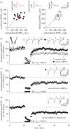
Tau is required for LTD in vivo in mice. (a) No differences are shown in synaptic transmission between MAPT+/+, MAPT+/− and MAPT−/− mice. The graph plots the fEPSP amplitude versus the fibre volley (f.v.) amplitude (stimulus intensity range: 10–100 μA, grey dots). Input-output curves show regression (continuous lines) and 95% confidence limits (dashed lines). (b) No differences are shown between MAPT+/+, MAPT+/− and MAPT−/− mice in paired-pulse facilitation at various inter-stimulus intervals. (c) LTD is absent in MAPT+/− and MAPT−/− mice. Pooled data from mice (age between 7 and 11 months; MAPT+/+: 76 ± 2%; n = 16; MAPT+/−: 96 ± 3%; n = 15; MAPT−/−: 98 ± 2%; n = 11; p < 0.001 in comparison with MAPT+/+ mice, Bonferroni's multiple comparison test). (d) No differences are shown in LTP between genotype. Pooled data from 7- to 11-month-old MAPT+/+ (141 ± 7% of baseline quantified at 60 min after the tetanus, n = 5), MAPT+/− (141 ± 6%, n = 6) and MAPT−/− mice (134 ± 3%, n = 4). HFS, high frequency stimulation. (Online version in colour.)
Figure 2.
Figure 2.
Tau is required for LTD in vitro in mice. (a) 1 Hz, 900 pulses induces LTD in MAPT+/+ mice (77 ± 3%, n = 6) but is absent in MAPT−/− mice (106 ± 7%, n = 5). Pooled data from postnatal 14- to 17-day-old mice. (b) Bath application of NMDA (25 μM, 3 min) induces LTD in MAPT+/+ mice (71 ± 8%, n = 5) but no LTD in MAPT−/− mice (99 ± 4%, n = 6). Pooled data from postnatal 14- to 17-day-old mice.
Figure 3.
Figure 3.
Knockdown of tau blocks LTD in rats. (a) Pairwise analysis of basal synaptic transmission between rat tau-shRNA expressing and untransfected neighbouring neurons, each obtained from independent slices. (b) Simultaneous dual-patch recordings were made from rat tau-shRNA-transfected and untransfected neighbouring cells. LFS was delivered (at time indicated by the bar) and LTD quantified 30 min later. (c) Data from scrambled tau-shRNA transfected cells. (d) Data from cells co-expressing rat tau-shRNA and human tau. Error bars indicate s.e.m. (Online version in colour.)
Figure 4.
Figure 4.
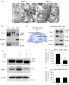
Tau is localized at the synapse and is phosphorylated during LTD. (a) Images of immunoelectron micrographs of hippocampal tissue obtained from MAPT+/+ (left panel; 4-month-old) and MAPT−/− (right panel; 4-month-old) mice. Arrow shows synaptic density and arrowheads indicate tau. JM (rabbit polyclonal anti-tau antibody) and 10 nm gold particle conjugated secondary antibody gave positive signals in MAPT+/+ but not in MAPT−/− mouse tissue. (b) Hippocampus of MAPT+/+ mouse (4 months old) was fractionated into a microsome/organelle fraction (P3), a cytoplasmic fraction (S3) and a PSD-95-rich fraction (LP1). MAP2 was mostly distributed in the P3 fraction. In comparison, tau (detected using Tau-5) was present in all fractions. (c) Schematic diagram of the microdissection procedure to separate the rat P24–28 CA1 somatic and dendritic regions. Western blotting shows strong expression of NeuN in the somatic region and of GluA2 in the dendritic region. Tau-5 blotting shows expression of tau in both the somatic and dendritic regions. (d) LFS causes an increase in phosphorylation of tau on Ser396/404 (PHF-1 epitope); this LFS-induced increase in phosphorylation of the PHF-1 epitope is attenuated by co-treatment with CT-99021 (CT; 1 μM). (e) Quantification of PHF-1 levels normalized to Tau-5 in the presence and absence of CT-99021 (control (CTR) versus LFS, *p < 0.05; LFS versus LFS + CT, **p < 0.01). (f) Quantification of Tau-5 expression, normalized to β-actin, in the presence and absence of CT-99021 (CTR versus LFS, p > 0.05; LFS versus LFS+CT, p > 0.05). Mann–Whitney non-parametric test was performed to identify changes in statistical significance. (Online version in colour.)

References

    1. Andreadis A, Brown WM, Kosik KS. 1992. Structure and novel exons of the human tau gene. Biochemistry 31, 10 626–10 633 (doi:10.1021/bi00158a027) - DOI - PubMed
    1. Goedert M, Wischik CM, Crowther RA, Walker JE, Klug A. 1988. Cloning and sequencing of the cDNA encoding a core protein of the paired helical filament of Alzheimer disease: identification as the microtubule-associated protein tau. Proc. Natl Acad. Sci. USA 85, 4051–4055 (doi:10.1073/pnas.85.11.4051) - DOI - PMC - PubMed
    1. Lindwall G, Cole RD. 1984. Phosphorylation affects the ability of tau protein to promote microtubule assembly. J. Biol. Chem. 259, 5301–5305 - PubMed
    1. Biernat J, Gustke N, Drewes G, Mandelkow EM, Mandelkow E. 1993. Phosphorylation of Ser262 strongly reduces binding of tau to microtubules: distinction between PHF-like immunoreactivity and microtubule binding. Neuron 11, 153–163 (doi:10.1016/0896-6273(93)90279-Z) - DOI - PubMed
    1. Weingarten MD, Lockwood AH, Hwo SY, Kirschner MW. 1975. A protein factor essential for microtubule assembly. Proc. Natl Acad. Sci. USA 72, 1858–1862 (doi:10.1073/pnas.72.5.1858) - DOI - PMC - PubMed

Publication types

MeSH terms

LinkOut - more resources