Review
. 2013 Dec 2;369(1633):20130284.
doi: 10.1098/rstb.2013.0284. Print 2014 Jan 5.

How the mechanisms of long-term synaptic potentiation and depression serve experience-dependent plasticity in primary visual cortex

Affiliations
Review

How the mechanisms of long-term synaptic potentiation and depression serve experience-dependent plasticity in primary visual cortex

Sam F Cooke et al. Philos Trans R Soc Lond B Biol Sci. .

Erratum in

  • Philos Trans R Soc Lond B Biol Sci. 2014 Apr 5;369(1639):20140021

Abstract

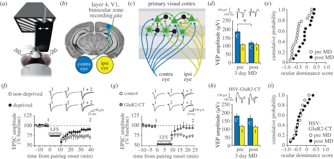
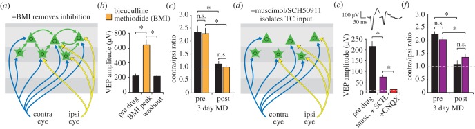
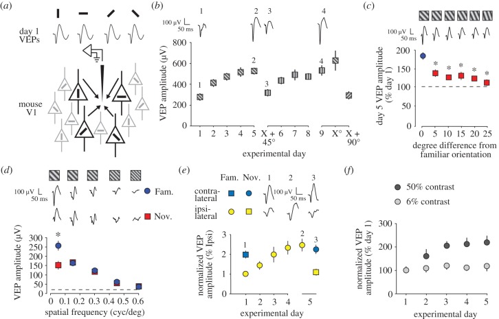
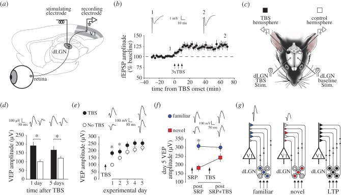
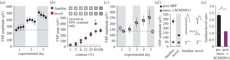
Donald Hebb chose visual learning in primary visual cortex (V1) of the rodent to exemplify his theories of how the brain stores information through long-lasting homosynaptic plasticity. Here, we revisit V1 to consider roles for bidirectional 'Hebbian' plasticity in the modification of vision through experience. First, we discuss the consequences of monocular deprivation (MD) in the mouse, which have been studied by many laboratories over many years, and the evidence that synaptic depression of excitatory input from the thalamus is a primary contributor to the loss of visual cortical responsiveness to stimuli viewed through the deprived eye. Second, we describe a less studied, but no less interesting form of plasticity in the visual cortex known as stimulus-selective response potentiation (SRP). SRP results in increases in the response of V1 to a visual stimulus through repeated viewing and bears all the hallmarks of perceptual learning. We describe evidence implicating an important role for potentiation of thalamo-cortical synapses in SRP. In addition, we present new data indicating that there are some features of this form of plasticity that cannot be fully accounted for by such feed-forward Hebbian plasticity, suggesting contributions from intra-cortical circuit components.

Keywords: amblyopia; long-term depression; long-term potentiation; monocular deprivation; perceptual learning; stimulus-selective response potentiation.


Figures

Figure 1.
Figure 1.
Ocular dominance (OD) plasticity resulting from visual deprivation. (a) Head-fixed mice view phase-reversing sinusoidal grating stimuli while visual-evoked potentials (VEPs) and unit activity are recorded. An occluder is used to restrict visual input to one eye or the other. (b) Recordings are made from electrodes implanted in layer 4 of the binocular zone of V1 (green), receiving independent input from the contralateral (blue) and ipsilateral (yellow) eyes. (c) In binocular V1 of the mouse, thalamo-recipient principal cells in layers 2/3 and 4 receive independent inputs from contralateral and ipsilateral eyes. Pronounced feed-forward connections from layer 4 to layer 2/3 and horizontal connections within layers 2/3 also exist. Inhibitory cells receive thalamic and intra-cortical input and inhibit principal cells throughout cortex. (d) OD plasticity is assayed by suturing the contralateral eye for 3 days to deprive this eye of visual input. Monocular deprivation (MD) results in a significant reduction in the amplitude of VEPs driven by visual input through the contralateral eye. Example waveforms are displayed at the top of the figure. (e) Binocularly responsive units can be scored for ocular dominance by assessing the bias towards response to the contralateral or ipsilateral eye prior to MD (open circles). After 3 days of MD this OD index shifts away from the deprived eye towards the non-deprived eye (closed circles). (f) Pairing of low-frequency stimulation (LFS) of white matter and layer 4 cell depolarization in V1 slices induces thalamo-cortical LTD (open circles). MD occludes this Hebbian LTD, preventing it from being established long-term (closed circles). Data are reproduced from [15]. (g) Clathrin-dependent endocytosis can be blocked by expression of a peptide that mimics the cytoplasmic tail of the GluR2 subunit (GluR2-CT). Expression of the GluR2-CT peptide blocks LFS pairing-induced Hebbian thalamocortical (TC) LTD in layer 4 (dark grey) of V1 slices relative to GFP-only control (light grey). (h) This same treatment prevents the OD shift resulting from MD relative to the interleaved controls presented in (d). (i) The ocular dominance shift resulting from MD is also blocked by the GluR2-CT peptide. This block can be compared to controls shown in (e). (d, e, g, h and i) are reproduced from [16]. Throughout the figure asterisks denote comparisons revealing significance of p < 0.05.
Figure 2.
Figure 2.
OD plasticity does not require inhibition for expression. (a) An in vivo pharmacological approach applies drugs locally around the VEP recording site in V1 in awake animals. Phasic inhibition can be blocked by applying the GABAA receptor antagonist bicuculline methiodide (BMI). (b) VEP amplitude is significantly enhanced as a result of BMI application (orange) but recovers after washout. (c) Measurement of VEPs driven independently through the two eyes reveals the approximately 2 : 1 ratio of the amplitude of contra- and ipsi-laterally driven VEPs, respectively. Despite the increase in VEP amplitude with BMI application this ratio is unchanged. After 3 days of MD, the contra : ipsi ratio falls significantly to approximately 1 : 1. Again this new ratio is unchanged in the presence of BMI, despite the overall increase in VEP amplitude, indicating a lack of requirement for modulated inhibition in the maintenance of the initial ratio and its shift as a result of MD. (d) Unit activity and, hence, all intra-cortical synaptic activity can be blocked through application of the GABAA receptor agonist muscimol. Thalamo-cortical input can be maintained also by locally infusing the GABAB receptor blocker SCH50911 to prevent non-specific muscimol binding pre-synaptically. (e) A TC VEP of significantly reduced amplitude can be isolated in the presence of the muscimol–SCH50911 cocktail (purple). This remaining VEP is completely abolished by the AMPAR antagonist CNQX (red), indicating that it is post-synaptically mediated. (f) The approximate 2 contra : 1 ipsi OD VEP ratio is maintained in the presence of this cocktail, as is expression of the shift as a result of 3 days of MD. (b, c, e and f) are reproduced from [57]. Asterisks denote significance of p < 0.05 while n.s. denotes higher p-values.
Figure 3.
Figure 3.
Stimulus-selective response potentiation (SRP). (a) In the rodent, unlike in cats, ferrets and primates, a confetti-like, interleaved arrangement of orientation selectivity exists in primary visual cortex (V1). As a consequence, visual evoked potentials (VEPs) of approximately equal trough-peak amplitude are driven by visual stimuli of any orientation at a single recording site within binocular V1 without prior modification through experience. (b) SRP occurs over days in awake mice as they view full-field sinusoidal grating stimuli. VEP amplitude undergoes potentiation through SRP that is selective for the orientation that has been experienced, as revealed by presenting a new orientation (in this case X + 45°). SRP then independently occurs to this new orientation at an equivalent rate and magnitude to that observed for X°. SRP can occur sequentially over multiple stimulus orientations. Example waveforms are shown at the top. (c) SRP is exquisitely orientation-selective so that the familiar orientation (blue) evokes significantly larger VEPs than a novel (red) orientation that is shifted by as little as 5°. (d) SRP is selective for the spatial frequency as well as the orientation of the stimulus. An oriented grating presented only at 0.05 cycles per degree (CPD) induces SRP (blue) that does not transfer across presentations of the same orientation at other spatial frequencies. This is revealed by comparisons to a novel orientation (red) presented across the same range of spatial frequencies because VEPs vary in amplitude depending on spatial frequency regardless of prior experience. (e) SRP can be induced through one eye only (in this case the ipsi-lateral eye in yellow) and this plasticity fails to transfer to the opposite eye (in this case the contra-lateral eye in blue). (f) SRP does not occur at low stimulus contrasts (grey) as compared to high contrasts within the same animals (black). (b, e and f) are reproduced from [86]. (c,d) are reproduced from [87]. Asterisks denote significance of p < 0.05.
Figure 4.
Figure 4.
SRP and LTP mutually occlude one another: (a) Geniculo-cortical LTP can be induced in vivo by acutely placing a stimulating electrode into the dorsal lateral geniculate nucleus (dLGN) of isoflurane anaesthetised mice and recording electrically-evoked potentials in layer 4 through chronically implanted recording electrodes that can also record VEPs in the same animals when awake. (b) LTP induced with theta burst stimulation (TBS) applied repeatedly to the dLGN lasts for at least the hour of recording time prior to recovery of animals from anaesthesia. (c) Interactions between LTP and SRP can be tested by inducing LTP in just one hemisphere (black) and comparing VEP amplitudes with those recorded in the other, control hemisphere (open) before or after SRP. (d) VEP amplitude was significantly greater in the LTP hemisphere (black) than the control hemisphere (open) whether VEPs were sampled 1 day later or 5 days later, indicating the impact of LTP on responsiveness to visual stimuli. (e) Although VEPs are significantly greater in amplitude in the TBS hemisphere than in the control hemisphere there is less SRP in this hemisphere as a consequence, so that VEP amplitude is no longer significantly different after 5 days of repeated presentation of the same grating stimulus. (f) In the reverse experiment, after SRP has been saturated to one familiar orientation (blue), TBS-induced LTP significantly impacts only those VEPs driven by a novel orientation (red), indicating that SRP occludes the effects of LTP on the VEP. (b–f) are reproduced from [87]. (g) A schematic describing the simple interpretation of these results shows that feed-forward TC plasticity forms templates of strengthened synapses for familiar stimuli (blue) in thalamo-recipient layer 4 that overlap minimally with patterns driven by other, novel oriented stimuli (red). LTP indiscriminately potentiates large numbers of synapses impacting response to all stimuli (black), whether familiar or novel. Asterisks denote significance of p < 0.05.
Figure 5.
Figure 5.
The complexity of SRP. (a) SRP does not emerge within the recording session as multiple samples within a half hour recording session yield VEPs of similar amplitude. Gains in amplitude are only apparent the following day. (b) SRP is selective for the contrast at which the stimulus is viewed. An oriented grating presented only at 50% contrast induces SRP (blue) that does not transfer across presentations of the same orientation at other contrasts, including the higher contrast of 100%. This is revealed by comparisons to a novel orientation (red) presented across the same range of contrasts because VEPs vary in amplitude depending on contrast regardless of prior experience. (c) SRP can occur to a compound chessboard stimulus. VEPs driven by the chessboard stimulus are of equivalent amplitude to those driven by a component orientation on day 1. However, SRP to the chessboard stimulus does not transfer to a second, novel component stimulus within an interleaved test session on day 5. (a–c) are reproduced from [86]. (d) Application of a cocktail of muscimol and SCH50911 (purple) isolates a purely TC VEP within layer 4 (as described in figure 2d). This treatment prevents expression of SRP such that VEPs driven by either familiar or novel stimuli are not significantly different in amplitude. Example VEPs are shown on the right side. (e) The ratio of VEP amplitude driven by familiar and novel stimuli falls significantly from approximately 2 : 1 prior to cocktail application to 1 : 1 after cocktail application, indicating that SRP is not simply expressed through potentiation of layer 4 TC synapses. (d) and (e) display new and previously unpublished data. Asterisks denote significance of p < 0.05.

References

    1. Bain A. 1873. Mind and body. The theories of their relation. New York, NY: D. Appleton and Company.
    1. Hebb DO. 1949. The organization of behavior. New York, NY: Wiley.
    1. Wiesel TN, Hubel DH. 1963. Single-cell responses in striate cortex of kittens deprived of vision in one eye. J. Neurophysiol. 26, 1003–1017. - PubMed
    1. Stent GS. 1973. A physiological mechanism for Hebb's postulate of learning. Proc. Natl Acad. Sci. USA 70, 997–1001. (10.1073/pnas.70.4.997) - DOI - PMC - PubMed
    1. Katz LC, Shatz CJ. 1996. Synaptic activity and the construction of cortical circuits. Science 274, 1133–1138. (10.1126/science.274.5290.1133) - DOI - PubMed

Publication types