

Signal and depth enhancement for in vivo flow cytometer measurement of ear skin by optical clearing agents
- PMID: 24298412
- PMCID: PMC3829546
- DOI: 10.1364/BOE.4.002518
Signal and depth enhancement for in vivo flow cytometer measurement of ear skin by optical clearing agents
Abstract
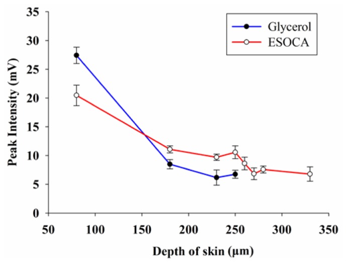
The in vivo flow cytometry (IVFC) has shown a great potential for detecting circulating tumor cells quantitatively in the bloodstream. However, the detection depth suffers from the strong light scattering of tissue. In this study, an innovative ear skin optical clearing agent (ESOCA) is employed to improve the signal quality of the IVFC. Our results show that compared with commonly used glycerol, topical application of ESOCA can enhance the transmittance of rat ear significantly in vivo. The labeled red blood cells can be detected by the IVFC with higher signal quality and greater detection depth. This study is very helpful for potential tumor metastasis studies by the IVFC in deep tissues.
Keywords: (170.1470) Blood or tissue constituent monitoring; (170.1530) Cell analysis; (170.3660) Light propagation in tissues.
Figures


Similar articles
-
Optical clearing of skin using flash lamp-induced enhancement of epidermal permeability.Lasers Surg Med. 2006 Oct;38(9):824-36. doi: 10.1002/lsm.20392. Lasers Surg Med. 2006. PMID: 17044094
-
Enhancement of skin optical clearing efficacy using photo-irradiation.Lasers Surg Med. 2010 Feb;42(2):132-40. doi: 10.1002/lsm.20900. Lasers Surg Med. 2010. PMID: 20166162
-
In Vivo Flow Cytometry.Adv Exp Med Biol. 2021;3233:289-305. doi: 10.1007/978-981-15-7627-0_13. Adv Exp Med Biol. 2021. PMID: 34053032
-
Advances of In Vivo Flow Cytometry on Cancer Studies.Cytometry A. 2020 Jan;97(1):15-23. doi: 10.1002/cyto.a.23851. Epub 2019 Jul 5. Cytometry A. 2020. PMID: 31273910 Review.
-
Optical clearing for multiscale biological tissues.J Biophotonics. 2018 Feb;11(2). doi: 10.1002/jbio.201700187. Epub 2017 Dec 12. J Biophotonics. 2018. PMID: 29024450 Review.
Cited by
-
Accessing to arteriovenous blood flow dynamics response using combined laser speckle contrast imaging and skin optical clearing.Biomed Opt Express. 2015 May 6;6(6):1977-89. doi: 10.1364/BOE.6.001977. eCollection 2015 Jun 1. Biomed Opt Express. 2015. PMID: 26114023 Free PMC article.
-
In vivo quantitation of injected circulating tumor cells from great saphenous vein based on video-rate confocal microscopy.Biomed Opt Express. 2015 May 19;6(6):2158-67. doi: 10.1364/BOE.6.002158. eCollection 2015 Jun 1. Biomed Opt Express. 2015. PMID: 26114035 Free PMC article.
-
In vivo cell characteristic extraction and identification by photoacoustic flow cytography.Biomed Opt Express. 2015 Sep 3;6(10):3748-56. doi: 10.1364/BOE.6.003748. eCollection 2015 Oct 1. Biomed Opt Express. 2015. PMID: 26504626 Free PMC article.
-
In Vivo Flow Cytometric Evaluation of Circulating Metastatic Pancreatic Tumor Cells after High-Intensity Focused Ultrasound Therapy.Cytometry A. 2020 Sep;97(9):900-908. doi: 10.1002/cyto.a.24014. Epub 2020 Apr 19. Cytometry A. 2020. PMID: 32307867 Free PMC article.
-
Optical clearing based cellular-level 3D visualization of intact lymph node cortex.Biomed Opt Express. 2015 Sep 28;6(10):4154-64. doi: 10.1364/BOE.6.004154. eCollection 2015 Oct 1. Biomed Opt Express. 2015. PMID: 26504662 Free PMC article.
References
-
- Terentyuk G. S., Maslyakova G. N., Suleymanova L. V., Khlebtsov N. G., Khlebtsov B. N., Akchurin G. G., Maksimova I. L., Tuchin V. V., “Laser-induced tissue hyperthermia mediated by gold nanoparticles: toward cancer phototherapy,” J. Biomed. Opt. 14(2), 021016 (2009).10.1117/1.3122371 - DOI - PubMed
-
- Herrmann A., Kortylewski M., Kujawski M., Zhang C., Reckamp K., Armstrong B., Wang L., Kowolik C., Deng J., Figlin R., Yu H., “Targeting Stat3 in the myeloid compartment drastically improves the in vivo antitumor functions of adoptively transferred T cells,” Cancer Res. 70(19), 7455–7464 (2010).10.1158/0008-5472.CAN-10-0736 - DOI - PMC - PubMed
LinkOut - more resources
Full Text Sources
Other Literature Sources