. 2013 Oct 24;4(11):2596-608.
doi: 10.1364/BOE.4.002596. eCollection 2013.

Automated lamina cribrosa microstructural segmentation in optical coherence tomography scans of healthy and glaucomatous eyes

Affiliations

Automated lamina cribrosa microstructural segmentation in optical coherence tomography scans of healthy and glaucomatous eyes

Zach Nadler et al. Biomed Opt Express. .

Abstract

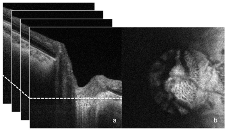
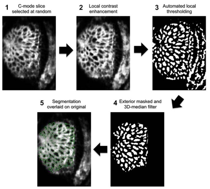
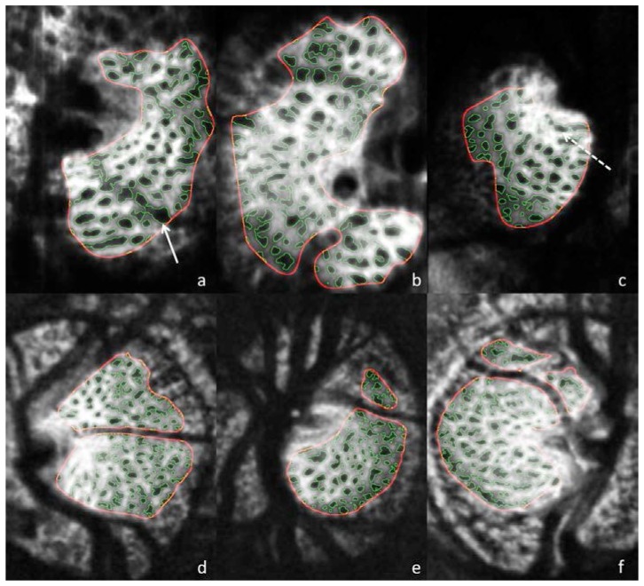
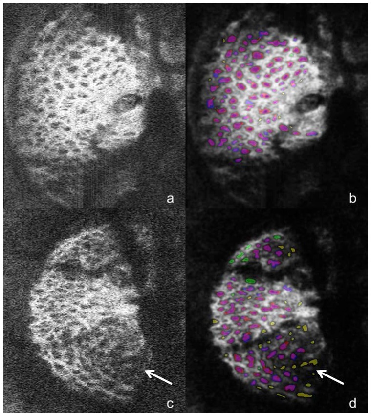
We demonstrate an automated segmentation method for in-vivo 3D optical coherence tomography (OCT) imaging of the lamina cribrosa (LC). Manual segmentations of coronal slices of the LC were used as a gold standard in parameter selection and evaluation of the automated technique. The method was validated using two prototype OCT devices; each had a subject cohort including both healthy and glaucomatous eyes. Automated segmentation of in-vivo 3D LC OCT microstructure performed comparably to manual segmentation and is useful for investigative research and in clinical quantification of the LC.

Keywords: (100.2000) Digital image processing; (110.4500) Optical coherence tomography; (170.1610) Clinical applications; (170.4470) Ophthalmology; (330.4460) Ophthalmic optics and devices.


Figures

Fig. 1
Fig. 1
SS-OCT scan of healthy eye. B-scan frames are stacked (a) into a 3D data cube from which an individual C-mode slice is selected at random (at the location of the dotted line on the left) to undergo manual and automated segmentation analysis (b).
Fig. 2
Fig. 2
Automated pore segmentation process for MAO-OCT scan. Following a 3D-Gaussian filter, a C-mode slice is randomly selected from the stack (1). A local contrast enhancement highlights local features of the structure (2), which are then thresholded (3). A 3D median filter removes pores unconnected in depth and the regions exterior to the visible lamina are masked (4). Finally, the segmentation is overlaid on the original image for subjective evaluation (5). The outline for automated segmentation is shown in green.
Fig. 3
Fig. 3
Automated segmentation results for MAO-OCT (a-c) and SS-OCT (d-f). Solid arrow (a) points to a region where blood vessel shadow is unmasked, and as a result the pore boundaries exhibit irregular boarders within the shadow. Dotted arrow (c) shows region where small adjacent pores are combined into a single pore.
Fig. 4
Fig. 4
The unprocessed C-mode slice (a) and corresponding segmentation (b) for a scan of a healthy eye taken with the SS-OCT device. For pores identified by both automated and manual segmentations the automated pores are colored red and manual are colored blue so that overlapping segmentation appears as purple. Pores identified only by the automated method are colored yellow and those seen solely in the manual segmentation are colored green.
Fig. 5
Fig. 5
The unprocessed C-mode slice (a,c) and corresponding segmentation (b,d) for scans of one glaucomatous (top) and one healthy eye (bottom) taken with the MAO-OCT system. Segmentations are colored according the scheme outlined in Fig. 4. The top image shows relatively good agreement between manual and automated methods, while the bottom images exhibit regions of segmentation disagreement; the arrow points to pores identified in the automated method but not manually. These locations exhibits low signal-to-noise ratio in the original image (c), which explains the discrepancy.

References

    1. Quigley H. A., Broman A. T., “The number of people with glaucoma worldwide in 2010 and 2020,” Br. J. Ophthalmol. 90(3), 262–267 (2006).10.1136/bjo.2005.081224 - DOI - PMC - PubMed
    1. Townsend K. A., Wollstein G., Schuman J. S., “Imaging of the retinal nerve fibre layer for glaucoma,” Br. J. Ophthalmol. 93(2), 139–143 (2009).10.1136/bjo.2008.145540 - DOI - PMC - PubMed
    1. Wollstein G., Schuman J. S., Price L. L., Aydin A., Stark P. C., Hertzmark E., Lai E., Ishikawa H., Mattox C., Fujimoto J. G., Paunescu L. A., “Optical coherence tomography longitudinal evaluation of retinal nerve fiber layer thickness in glaucoma,” Arch. Ophthalmol. 123(4), 464–470 (2005).10.1001/archopht.123.4.464 - DOI - PMC - PubMed
    1. Leung C. K., Choi N., Weinreb R. N., Liu S., Ye C., Liu L., Lai G. W., Lau J., Lam D. S., “Retinal nerve fiber layer imaging with spectral-domain optical coherence tomography: pattern of RNFL defects in glaucoma,” Ophthalmology 117(12), 2337–2344 (2010).10.1016/j.ophtha.2010.04.002 - DOI - PubMed
    1. Tan O., Li G., Lu A. T., Varma R., Huang D., Advanced Imaging for Glaucoma Study Group , “Mapping of macular substructures with optical coherence tomography for glaucoma diagnosis,” Ophthalmology 115(6), 949–956 (2008).10.1016/j.ophtha.2007.08.011 - DOI - PMC - PubMed

LinkOut - more resources