. 2013;15(6):R207.
doi: 10.1186/ar4400.

PDL241, a novel humanized monoclonal antibody, reveals CD319 as a therapeutic target for rheumatoid arthritis

PDL241, a novel humanized monoclonal antibody, reveals CD319 as a therapeutic target for rheumatoid arthritis

Jacky Woo et al. Arthritis Res Ther. 2013.

Abstract

Introduction: Targeting the CD20 antigen has been a successful therapeutic intervention in the treatment of rheumatoid arthritis (RA). However, in some patients with an inadequate response to anti-CD20 therapy, a persistence of CD20- plasmablasts is noted. The strong expression of CD319 on CD20- plasmablast and plasma cell populations in RA synovium led to the investigation of the potential of CD319 as a therapeutic target.

Methods: PDL241, a novel humanized IgG1 monoclonal antibody (mAb) to CD319, was generated and examined for its ability to inhibit immunoglobulin production from plasmablasts and plasma cells generated from peripheral blood mononuclear cells (PBMC) in the presence and absence of RA synovial fibroblasts (RA-SF). The in vivo activity of PDL241 was determined in a human PBMC transfer into NOD scid IL-2 gamma chain knockout (NSG) mouse model. Finally, the ability of PDL241 to ameliorate experimental arthritis was evaluated in a collagen-induced arthritis (CIA) model in rhesus monkeys.

Results: PDL241 bound to plasmablasts and plasma cells but not naïve B cells. Consistent with the binding profile, PDL241 inhibited the production of IgM from in vitro PBMC cultures by the depletion of CD319+ plasmablasts and plasma cells but not B cells. The activity of PDL241 was dependent on an intact Fc portion of the IgG1 and mediated predominantly by natural killer cells. Inhibition of IgM production was also observed in the human PBMC transfer to NSG mouse model. Treatment of rhesus monkeys in a CIA model with PDL241 led to a significant inhibition of anti-collagen IgG and IgM antibodies. A beneficial effect on joint related parameters, including bone remodeling, histopathology, and joint swelling was also observed.

Conclusions: The activity of PDL241 in both in vitro and in vivo models highlights the potential of CD319 as a therapeutic target in RA.


Figures

Figure 1
Figure 1
CD319 is expressed on RA synovial tissue (IHC) and is a marker of plasma cells. FFPE samples of synovial tissues from RA patients were used for IHC analysis of CD319 using (A) 1G9 alone and co-stained with cell surface markers for leukocyte subsets (B-F). Double labeling studies were performed using 1G9 in combination with (B) anti-CD3 (T cells), (C) anti-CD20 (B cells), (D) anti-CD56 (NK cells), (E) anti-CD68 (macrophages), and (F) CD138 for plasma cells. Brown staining represented CD319 reactivity while other cell surface markers stained red. Double staining cells are purple. FFPE, formalin fixed paraffin embedded; IHC, immunohistochemistry; RA, rheumatoid arthritis.
Figure 2
Figure 2
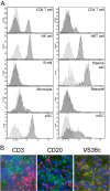
Binding of PDL241 to leukocytes. (A) PBMC subsets were labeled for immunofluorescence analysis on the basis of co-staining: CD3+ CD4+ and CD3 + CD8+ (T cells); CD3-CD56+ (NK cells) and CD3 + CD56+ (NKT cells); CD19+ (B cells) and CD14+ (monocytes); CD27 + CD38 + CD138+ (plasmablasts and plasma cells). Lineage negative cells were first gated and then separated into HLA-DR + CD11c + (mDC), HLA-DR + CD123+ (pDC) and HLA-DR-CD123+ (basophils). Data representative of 1 of 10 donors. (B)  Immunofluorescence analysis of co-staining of PDL241 (green) with anti-CD3 (T cells), anti-CD20 (B cells) or VS38c Ab (plasma cells) in red on OCT embedded frozen tissues from normal human tonsil. Nuclei were counter-stained with DAPI (blue). Double stained cells appeared yellow. Ab, antibody; DAPI, 4′,6-diamidino-2-phenylindole; NK, natural killer; OCT, optimal cutting temperature; PBMC, peripheral blood mononuclear cells.
Figure 3
Figure 3
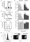
PDL241 inhibits Ig production from PBMC by specifically depleting plasmablasts and plasma cells. (A) Time course of inhibition of IgM production from PBMC. Representative of n = 2 experiments. (B) Inhibition of IgM by PDL241 is dependent on mAb concentration and intact mAb, as F(ab’)2 fragments of PDL241 showed no activity. The dose dependent inhibition of IgM by PDL241 was demonstrated in >10 donors; F(ab’)2 experiment was a representative experiment of two. (C) Depletion of NK cells but not monocytes from PBMC reduces the inhibitory activity of PDL241. Representative experiment of n = 4; P = 0.01 for NK cell depleted compared to PBMC. (D) PDL241 treatment results in specific reduction in plasmablast counts in PBMC cultures. In contrast to rituximab (open bars), PDL241 treatment (closed bars) of PBMC specifically depleted plasma cells but not B cells from PBMC cultures. Counts were expressed as% of cIgG1-treated cultures. Data represent the mean and SD of n = 6 experiments from distinct donors. P ≤0.001 for B cell depletion by rituxan compared to PDL241 at 1 and 10 μg/ml. (E) CD319 was highly expressed on plasmablasts in PBMC cultures, (left panel, black histogram). Dot plots showing CD27 + CD38+ plasmablasts following treatment with cIgG1 control (middle panel) or PDL241 (right panel). Ig, immunoglobulin; mAB, monoclonal antibody; NK, natural killer; PBMC, peripheral blood mononuclear cells; SD, standard deviation.
Figure 4
Figure 4
PDL241 treatment of RA synovial fibroblast-PBMC co-cultures leads to reduction on plasmablasts with no effect on B cells. (A) B cells differentiated into CD19+CD27hiCD38hiCD319+ plasmablasts after co-culture with RA-synovial fibroblasts. PBMC from normal donors were cultured with RA synovial fibroblasts. At day 0 and day 7, cells in the culture were analyzed by flow cytometry. After co-culture for seven days, differentiated plasmablast cells (CD19+CD27hiCD38hi) (gate 1) could be detected and were found to be CD319 positive (solid line) as compared to isotype control staining (dotted line); in contrast, memory (CD19+CD27+) (gate 2) and naïve B cells (CD19+CD27) (gate 3) did not express CD319. (B) Removal of CD319+ plasmablast cells by PDL241 in RA synovial fibroblast-PBMC co-cultures. Addition of PDL241 to cultures specifically depleted CD19+CD27hiCD38hi plasmablasts, as compared to rituximab, which depleted only B cells. A FcR-binding deficient mutant of PDL241 (241-G2M3) had no effect on plasmablast cell numbers. The number in the gated population was calculated based on 10,000 events recoded. A representative experiment of n = 4 is shown. PBMC, peripheral blood mononuclear cells; RA, rheumatoic arthritis.
Figure 5
Figure 5
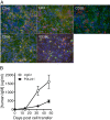
PDL241 binds human leukocytes and inhibits human Ig production in huSCID mice. NSG mice were transfused with human PBMC. (A) Double staining of CD319 (green fluorescence) and human leukocyte cell surface markers as indicated (red fluorescence) in the spleen. Cells expressing both markers were co-stained and appear yellow. 600X magnification. (B) Mice were sorted into groups on day 10 post-transfusion, and dosed twice weekly with PDL241 or cIgG1. IgM levels in the serum were measured at day 32 and day 47. In this experiment, PDL241 treatment resulted in significant reduction of human IgM compared to cIgG1 (day 32 P = 0.003 and day 47 P = 0.004 two tailed t-test). Significant reduction in IgM was observed in 6 of 11 separate experiments. huSCID, human-severe combined immunodeficiency; Ig, immunoglobulin; PBMC, peripheral blood mononuclear cells.
Figure 6
Figure 6
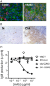
PDL241 binds to plasma cells in rhesus monkey. (A) Immunofluorescence co-staining of PDL241 and anti-CD20 or VS38c in tonsils from normal rhesus monkey. PDL241 staining was green, CD20 and VS38c stained red and nuclei were counter-stained with DAPI (blue). Cells with double staining (PDL241 and VS38c) appeared yellow. (B) IHC staining of PDL241 to draining lymph nodes from normal (N) or collagen immunized (CIA) rhesus monkey. (C). PDL241 but not Fc mutant 241-G2M3 inhibited IgM production from rhesus monkey PBMC stimulated with the TLR-9 agonist ODN2006. CIA, collagen-induced arthritis; DAPI, 4′,6-diamidino-2-phenylindole; IgM, immunoglobulin M; IHC, immunohistochemistry; PBMC, peripheral blood mononuclear cells.
Figure 7
Figure 7
PDL241 had no effect on clinical parameters in the rhesus monkey CIA model. The analysis of all animals showed no effect of PDL241 on (A) bodyweight loss relative to day 0; (B) average clinical score; and (C) serum CRP levels. PDL241 treatment had no effect on (D) disease free survival or (E) overall survival. CIA, collagen-induced arthritis; CRP, C-reactive protein.
Figure 8
Figure 8
PDL241 treatment reduced the severity of joint-related disease parameters in a rhesus CIA model. Analyses are provided for ‘all animals’ and animals with ‘early CRP onset’ as described in the Methods section. Collagen type II-specific IgM (A) and IgG (B) were measured by ELISA. (C) The small joint swelling score is a representation of the number of joints affected (joints with soft tissues swelling) and the severity of swelling for each individual joint. Urinary excretion of the collagen crosslinks (D) hydroxylysylpyrridinoline (HP) and (E) lysylpyrridinoline (LP) was determined, with the levels normalized to creatinine levels (nmol levels per mmol creatinine) to compensate for a possible dilution by spilled drinking water. Pl = Placebo, 30 = 30 mg/kg and 100 = 100 mg/kg. * P ≤0.05; ** P ≤0.01. CIA, collagen-induced arthritis; CRP, C-reactive protein; Ig, immunoglobulin.
Figure 9
Figure 9
PDL241 treatment reduced the severity of joint-related histopathological parameters in the rhesus CIA model. The histopathology score was based on a grading system described by Petit et al. [29]. Analyses were performed for (A) ‘all animal’ or (B) the ‘early CRP onset’ subgroup. CIA, collagen-induced arthritis; CRP, C-reactive protein.

Similar articles

Cited by

References

    1. McInnes IB, Schett G. The pathogenesis of rheumatoid arthritis. N Engl J Med. 2011;15:2205–2219. doi: 10.1056/NEJMra1004965. - DOI - PubMed
    1. Vander Cruyssen B, Peene I, Cantaert T, Hoffman IE, De Rycke L, Veys EM, De Keyser F. Anti-citrullinated protein/peptide antibodies (ACPA) in rheumatoid arthritis: specificity and relation with rheumatoid factor. Autoimmun Rev. 2005;15:468–474. doi: 10.1016/j.autrev.2005.04.018. - DOI - PubMed
    1. Genovese MC, Becker JC, Schiff M, Luggen M, Sherrer Y, Kremer J, Birbara C, Box J, Natarajan K, Nuamah I, Li T, Aranda R, Hagerty DT, Dougados M. Abatacept for rheumatoid arthritis refractory to tumor necrosis factor alpha inhibition. N Engl J Med. 2005;15:1114–1123. doi: 10.1056/NEJMoa050524. - DOI - PubMed
    1. Edwards JC, Szczepanski L, Szechinski J, Filipowicz-Sosnowska A, Emery P, Close DR, Stevens RM, Shaw T. Efficacy of B-cell-targeted therapy with rituximab in patients with rheumatoid arthritis. N Engl J Med. 2004;15:2572–2581. doi: 10.1056/NEJMoa032534. - DOI - PubMed
    1. Martin F, Chan AC. Pathogenic roles of B cells in human autoimmunity; insights from the clinic. Immunity. 2004;15:517–527. doi: 10.1016/S1074-7613(04)00112-8. - DOI - PubMed

Substances